 Back
The Nervous System: Structure, Function, and Physiology
Study Guide - Smart Notes
 Tailored notes based on your materials, expanded with key definitions, examples, and context.
Introduction to the Nervous System
Overview and Functions
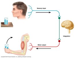
The nervous system is a highly complex organ system responsible for regulating and coordinating body activities. It works closely with the endocrine system to maintain homeostasis by monitoring internal and external environments, integrating information, and coordinating responses through effectors such as muscles and glands.
Monitoring: Constantly senses changes in the environment.
Integration: Processes and interprets sensory input.
Response: Initiates actions by sending signals to effectors.
Divisions of the Nervous System
Central and Peripheral Nervous Systems
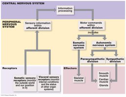
The nervous system is divided into two main parts:
Central Nervous System (CNS): Consists of the brain and spinal cord, responsible for processing and integrating information.
Peripheral Nervous System (PNS): Composed of all neural tissue outside the CNS, including nerves, ganglia, and receptors. It connects the CNS to limbs and organs.
Sensory and Motor Divisions of the PNS
Sensory (Afferent) Division: Transmits sensory information from receptors to the CNS.
Motor (Efferent) Division: Transmits commands from the CNS to effectors (muscles and glands).
Subdivisions of the Sensory and Motor Divisions
Somatic Sensory: Receives information from skin, skeletal muscles, and bones.
Visceral Sensory: Receives information from internal organs (viscera).
Somatic Motor: Controls voluntary movements of skeletal muscles.
Visceral Motor (Autonomic Nervous System, ANS): Controls involuntary actions of cardiac muscle, smooth muscle, and glands. Subdivided into sympathetic and parasympathetic divisions.
Cell Types in Nervous Tissue
Neurons and Neuroglia
Nervous tissue contains two main cell types:
Neurons: The functional units of the nervous system, specialized for communication. They do not divide after development.
Neuroglia (Glial Cells): Support, protect, and nourish neurons. They are more numerous than neurons and can divide throughout life.
Structure and Classification of Neurons
Basic Structure of a Neuron
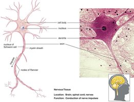
Soma (Cell Body): Contains the nucleus and organelles.
Dendrites: Receive incoming signals.
Axon Hillock: Site of action potential initiation.
Axon: Conducts electrical impulses away from the soma.
Axon Terminals: Communicate with target cells.
Structural Classification of Neurons
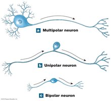
Multipolar Neuron: Multiple dendrites, one axon; most common type.
Unipolar Neuron: Single process extending from the soma; sensory neurons.
Bipolar Neuron: One dendrite and one axon; rare, found in special senses.
Functional Classification of Neurons
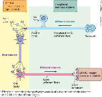
Afferent (Sensory) Neurons: Transmit impulses from receptors to the CNS; usually unipolar.
Interneurons: Located entirely within the CNS; process and relay information.
Efferent (Motor) Neurons: Transmit impulses from the CNS to effectors; usually multipolar.
Neuroglia (Glial Cells)
Neuroglia in the CNS
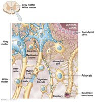
Astrocytes: Maintain the blood-brain barrier, provide structural support, and regulate the environment around neurons.
Oligodendrocytes: Form myelin sheaths around axons in the CNS, increasing the speed of electrical conduction.
Microglia: Act as phagocytes, removing debris and pathogens.
Ependymal Cells: Line brain ventricles and spinal cord central canal; produce and circulate cerebrospinal fluid (CSF).
Neuroglia in the PNS
Satellite Cells: Surround neuron cell bodies in the PNS, regulating the environment.
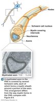
Schwann Cells: Form myelin sheaths around axons in the PNS and assist in axonal regeneration.
White and Gray Matter
Distribution in the CNS
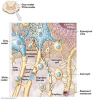
White Matter: Regions with myelinated axons; appears white due to lipid content of myelin.
Gray Matter: Regions with neuron cell bodies, dendrites, and unmyelinated axons; appears gray.
Electrical Properties of Neurons
Resting Membrane Potential (RMP)
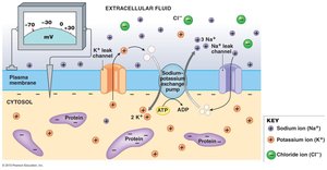
Neurons are polarized, with a typical RMP of -70 mV. This potential is established by:
Negatively charged proteins inside the cell.
More potassium (K+) leak channels than sodium (Na+) channels, leading to more K+ leaving the cell.
The sodium-potassium pump, which moves 3 Na+ out and 2 K+ in, maintaining the gradient.
Action Potentials
Generation and Phases
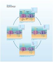
An action potential is a rapid, large change in membrane potential that travels along the axon. It is initiated at the axon hillock and involves voltage-gated Na+ and K+ channels.
Depolarization: Voltage-gated Na+ channels open rapidly, Na+ influxes, membrane potential becomes positive.
Repolarization: Voltage-gated K+ channels open slowly, K+ effluxes, membrane potential returns to negative.
Hyperpolarization: Membrane potential becomes more negative than RMP before stabilizing.
Propagation of Action Potentials
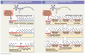
Contiguous (Continuous) Propagation: Occurs in unmyelinated axons; action potential moves along every segment of the membrane.
Saltatory Propagation: Occurs in myelinated axons; action potential jumps from node of Ranvier to node of Ranvier, increasing speed.
Synapses
Chemical Synapses and Transmission
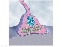
A synapse is the junction between two neurons. Most synapses in the nervous system are chemical synapses, where neurotransmitters mediate communication.
Presynaptic Neuron: Releases neurotransmitter from synaptic vesicles.
Synaptic Cleft: Small gap between neurons.
Postsynaptic Neuron: Receives neurotransmitter, leading to excitation or inhibition.
Events at a Cholinergic Synapse
An action potential arrives at the axon terminal, depolarizing the membrane.
Voltage-gated Ca2+ channels open, Ca2+ enters, triggering exocytosis of acetylcholine (ACh).
ACh diffuses across the synaptic cleft and binds to receptors on the postsynaptic membrane, opening ligand-gated Na+ channels and causing depolarization.
ACh is degraded by acetylcholinesterase (AChE), terminating the signal.
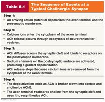
Summary Table: Events at a Cholinergic Synapse
Step | Description |
|---|---|
1 | Action potential depolarizes axon terminal and presynaptic membrane. |
2 | Ca2+ enters axon terminal, triggering exocytosis of neurotransmitter vesicles. |
3 | ACh diffuses across synaptic cleft, binds to postsynaptic receptors, and opens Na+ channels. |
4 | Na+ influx causes depolarization of postsynaptic membrane. |
5 | ACh is degraded by AChE, ending the signal. |
