 Back
The Nervous System: Structure, Function, and Organization
Study Guide - Smart Notes
 Tailored notes based on your materials, expanded with key definitions, examples, and context.
The Nervous System: Overview and Functions
Introduction to the Nervous System
The nervous system is the master control and communication system of the body, responsible for regulating every thought, action, and emotion. It communicates with body cells using rapid and specific electrical impulses, resulting in immediate responses. The nervous system works closely with the endocrine system to maintain homeostasis, but it uses electrical signals while the endocrine system uses hormones released into the blood.
Key Functions: Sensory input, integration, and motor output.
Feedback Loop: Sensory receptors detect stimuli, the brain integrates information, and effectors (muscles/glands) respond.
Structural and Functional Organization
Central and Peripheral Nervous Systems
The nervous system is structurally divided into the central nervous system (CNS) and the peripheral nervous system (PNS).
CNS: Consists of the brain and spinal cord; acts as the integrating and command center.
PNS: Includes all nerves outside the CNS; connects the CNS to limbs and organs.
Functional Divisions of the PNS
Sensory (Afferent) Division: Transmits impulses from sensory receptors to the CNS.
Motor (Efferent) Division: Transmits impulses from the CNS to effectors (muscles/glands).
Somatic Nervous System: Controls voluntary movements of skeletal muscles.
Autonomic Nervous System (ANS): Controls involuntary functions (smooth muscle, cardiac muscle, glands); subdivided into sympathetic and parasympathetic divisions.
Nervous Tissue: Cells and Structure
Supporting Cells (Neuroglia)
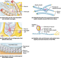
Neuroglia are supporting cells that protect, insulate, and support neurons. They do not conduct nerve impulses and retain the ability to divide, making them the source of most brain tumors (gliomas).
Astrocytes: Star-shaped cells that anchor neurons to capillaries and regulate the chemical environment.
Microglia: Phagocytic cells that remove debris and pathogens.
Ependymal Cells: Line brain and spinal cord cavities; circulate cerebrospinal fluid (CSF).
Oligodendrocytes: Form myelin sheaths around CNS nerve fibers.
Schwann Cells (PNS): Form myelin sheaths around PNS nerve fibers.
Satellite Cells (PNS): Cushion and protect neuron cell bodies in the PNS.
Neurons (Nerve Cells)
Neurons are specialized cells that transmit nerve impulses. They consist of a cell body (with nucleus), dendrites (receive signals), and a single axon (sends signals).
Cell Body: Contains nucleus and metabolic machinery; lacks centrioles (amitotic).
Dendrites: Receive incoming signals toward the cell body.
Axon: Conducts impulses away from the cell body; ends in axon terminals containing neurotransmitters.
Myelin Sheath: Fatty covering that insulates axons and speeds impulse transmission; formed by Schwann cells in PNS and oligodendrocytes in CNS.
Nodes of Ranvier: Gaps in the myelin sheath that facilitate rapid impulse conduction (saltatory conduction).
Neuronal Classification
Structural Classification
Multipolar Neurons: Many processes; most common, includes motor and interneurons.
Bipolar Neurons: One axon and one dendrite; rare, found in special sense organs.
Unipolar Neurons: Single process that divides; sensory neurons in PNS ganglia.
Functional Classification
Sensory (Afferent) Neurons: Carry impulses from receptors to CNS.
Motor (Efferent) Neurons: Carry impulses from CNS to effectors.
Interneurons (Association Neurons): Connect sensory and motor neurons within the CNS.
Nerve Impulse Transmission
Resting Membrane Potential and Action Potential
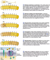
The resting neuron membrane is polarized, with more positive ions outside (Na+) than inside (K+). When stimulated, sodium channels open, Na+ enters, causing depolarization. If threshold is reached, an action potential (nerve impulse) is generated and propagated along the axon. Repolarization follows as K+ exits, restoring the resting state. The sodium-potassium pump restores ionic balance.
Saltatory Conduction: In myelinated fibers, impulses jump from node to node, increasing speed.
Synapse: The junction between neurons where neurotransmitters transmit signals across the synaptic cleft.
Reflexes and Reflex Arcs
Types and Components of Reflexes
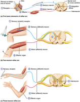
Reflexes are rapid, predictable, involuntary responses to stimuli. They occur over neural pathways called reflex arcs, which involve both CNS and PNS structures.
Somatic Reflexes: Stimulate skeletal muscles (e.g., withdrawal from pain).
Autonomic Reflexes: Regulate smooth muscle, cardiac muscle, and glands (e.g., pupillary reflex).
Reflex Arc Elements: Receptor, sensory neuron, integration center, motor neuron, effector.
Central Nervous System: Brain and Spinal Cord
Major Brain Regions and Functions
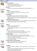
The brain consists of the cerebrum, diencephalon, brain stem, and cerebellum. Each region has specialized functions essential for sensory processing, motor control, and homeostasis.
Region | Major Functions |
|---|---|
Cerebral Cortex | Conscious thought, voluntary movement, sensory perception |
Basal Nuclei | Regulate voluntary motor activities |
Thalamus | Relay station for sensory impulses |
Hypothalamus | Homeostasis, emotion, autonomic control, endocrine regulation |
Epithalamus | CSF production, pineal gland (endocrine) |
Midbrain | Visual/auditory reflexes, motor tracts |
Pons | Breathing control, bridge for tracts |
Medulla Oblongata | Vital centers: heart rate, breathing, swallowing, vomiting |
Cerebellum | Coordination, balance, timing of movements |
Cerebral Cortex: Sensory and Motor Maps
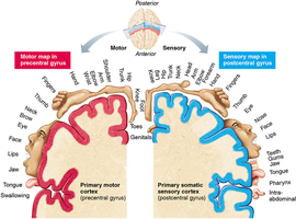
The cerebral cortex contains distinct areas for sensory and motor functions. The primary somatic sensory cortex (postcentral gyrus) receives sensory input, while the primary motor cortex (precentral gyrus) controls voluntary movements. The sensory and motor homunculi illustrate the amount of cortex devoted to different body regions.
Protection of the CNS
Meninges, Cerebrospinal Fluid, and Blood-Brain Barrier
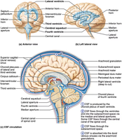
The CNS is protected by three connective tissue membranes (meninges), cerebrospinal fluid (CSF), and the blood-brain barrier.
Meninges: Dura mater (outer), arachnoid mater (middle), pia mater (inner).
CSF: Produced by choroid plexuses; circulates in ventricles and subarachnoid space, providing cushioning and nutrient transport.
Blood-Brain Barrier: Selective permeability protects brain from harmful substances.
Spinal Cord Structure and Function
Spinal Cord Anatomy
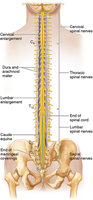
The spinal cord is a two-way conduction pathway between the brain and body and a major reflex center. It is protected by vertebrae and meninges, and ends at the level of the first or second lumbar vertebra.
Gray Matter: Butterfly-shaped; contains interneurons and motor neuron cell bodies.
White Matter: Myelinated fiber tracts; divided into dorsal, lateral, and ventral columns.
Spinal Nerves: 31 pairs; arise from fusion of dorsal and ventral roots.
Peripheral Nervous System (PNS)
Nerves and Ganglia
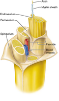
Nerves are bundles of neuron fibers outside the CNS, protected by connective tissue layers: endoneurium, perineurium, and epineurium. Ganglia are clusters of neuron cell bodies in the PNS.
Plexuses and Major Peripheral Nerves
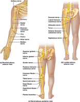
The ventral rami of spinal nerves (except T1-T12) form plexuses that serve the limbs. Major plexuses include the brachial, lumbar, and sacral plexuses, which give rise to important peripheral nerves.
Autonomic Nervous System (ANS)
Divisions and Functions
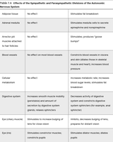
The ANS regulates involuntary activities of smooth muscle, cardiac muscle, and glands. It has two antagonistic divisions:
Sympathetic Division: Mobilizes body during stress ("fight or flight").
Parasympathetic Division: Conserves energy, promotes "rest and digest" activities.
Organ/System | Sympathetic Effect | Parasympathetic Effect |
|---|---|---|
Heart | Increases rate and force | Decreases rate |
Digestive System | Decreases activity | Increases activity |
Bronchioles | Dilates | Constricts |
Pupils | Dilates | Constricts |
Blood Vessels | Constriction (most) | Little/no effect |
Development and Aging of the Nervous System
Developmental Aspects
The nervous system forms early in embryonic development and continues to mature through childhood, primarily through myelination. Aging leads to gradual loss of neurons and decreased efficiency, especially in the sympathetic division. Circulatory problems can accelerate nervous system decline, but intellectual activity can be maintained with proper health care.
Additional info: The notes above include expanded academic context and examples for clarity and completeness, as required for a mini-textbook style study guide.
