 Back
BackThe Nervous System: Structure, Function, and Cellular Organization
Study Guide - Smart Notes
 Tailored notes based on your materials, expanded with key definitions, examples, and context.
The Nervous System: Overview and Divisions
Main Divisions of the Nervous System
The nervous system is the master control and communication system of the body. It is divided into two main parts: the Central Nervous System (CNS) and the Peripheral Nervous System (PNS).
Central Nervous System (CNS): Consists of the brain and spinal cord. It is responsible for integrating, processing, and coordinating sensory data and motor commands.
Peripheral Nervous System (PNS): Composed of nerves that extend from the brain and spinal cord to the rest of the body. It is further divided into the Somatic Nervous System (voluntary control of skeletal muscles) and the Autonomic Nervous System (involuntary control of smooth muscle, cardiac muscle, and glands).
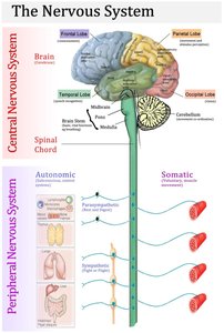
Divisions of the Peripheral Nervous System
Somatic Nervous System: Controls voluntary movements by innervating skeletal muscles.
Autonomic Nervous System (ANS): Regulates involuntary functions such as heart rate, digestion, and respiratory rate. The ANS is further divided into:
Sympathetic Division: Prepares the body for 'fight or flight' responses.
Parasympathetic Division: Promotes 'rest and digest' activities.
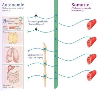
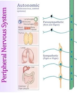
Functional Organization of the Nervous System
How Information Travels in the Nervous System
Information is transmitted through the nervous system via specialized cells called neurons. The flow of information follows a specific pathway:
Sensory (Afferent) Neurons: Detect changes inside and outside the body and transmit sensory information to the CNS.
Interneurons: Located within the CNS, these neurons process and integrate sensory input and determine appropriate responses.
Motor (Efferent) Neurons: Carry commands from the CNS to effectors such as muscles and glands.
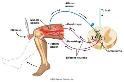
Neural Tissue: Structure and Function
Basic Structure of a Neuron
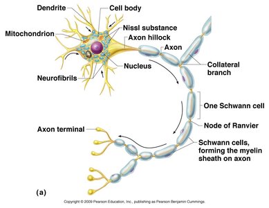
Neurons are the functional units of the nervous system. Each neuron consists of several key parts:
Dendrites: Highly branched extensions that receive electrical impulses and convey them toward the cell body.
Cell Body (Soma): Contains the nucleus and organelles; integrates incoming signals.
Axon: A single, long process that transmits impulses away from the cell body to other neurons or effectors.
Axon Hillock: The region where the axon originates from the cell body; site of action potential initiation.
Axon Terminals: Specialized endings that form synapses with other cells.
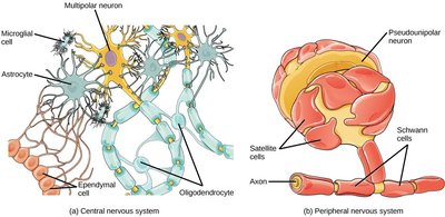
Schwann Cells: Glial cells in the PNS that produce myelin sheaths around axons.
Nodes of Ranvier: Gaps between Schwann cells where action potentials are regenerated.
Types of Neurons
Structural Classification
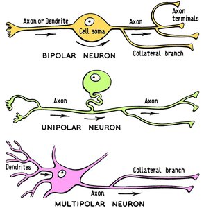
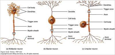
Neurons are classified based on the number and arrangement of their processes:
Bipolar Neurons: Have one axon and one dendrite; found in sensory organs such as the retina and olfactory epithelium.
Unipolar Neurons: Have a single process that splits into two branches; common in sensory neurons of the PNS.
Multipolar Neurons: Have one axon and multiple dendrites; the most common type in the CNS.
Neuroglia: Supporting Cells of the Nervous System
Types and Functions of Neuroglia
Neuroglia are non-neuronal cells that provide structural and functional support to neurons. They are classified based on their location:
Central Nervous System (CNS) | Peripheral Nervous System (PNS) |
|---|---|
Astrocytes: Regulate the blood-brain barrier, ion balance, and repair after injury. Oligodendrocytes: Myelinate axons in the CNS. Microglia: Act as phagocytes, removing debris and pathogens. Ependymal Cells: Line ventricles and produce cerebrospinal fluid. | Schwann Cells: Myelinate axons in the PNS. Satellite Cells: Support neuron cell bodies in ganglia. |
Membrane Potentials and Ion Channels
Resting Membrane Potential
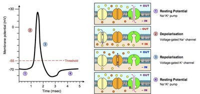
The cell membrane of a neuron is polarized due to the unequal distribution of ions, primarily maintained by the sodium-potassium pump. The typical resting membrane potential is -70 mV.
Na+/K+ Pump: Actively transports 3 Na+ ions out and 2 K+ ions into the cell, contributing to the negative charge inside the neuron.
Types of Ion Channels
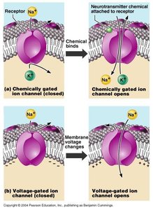
Leakage (Nongated) Channels: Always open; contribute to resting potential.
Chemically Gated (Ligand-Gated) Channels: Open in response to binding of a chemical messenger.
Voltage-Gated Channels: Open or close in response to changes in membrane potential.
Mechanically Gated Channels: Open in response to physical deformation of the membrane.
Action Potentials and Signal Transmission
Generation and Propagation of Action Potentials
An action potential (AP) is a rapid change in membrane potential that travels along the axon. The process involves several phases:
Resting Potential: The neuron is at -70 mV, maintained by the Na+/K+ pump.
Depolarization: Voltage-gated Na+ channels open, causing Na+ influx and the membrane potential to become less negative.
Repolarization: Voltage-gated K+ channels open, K+ exits the cell, restoring negativity.
Hyperpolarization: Membrane potential temporarily becomes more negative than resting potential.
Refractory Period: The neuron cannot fire another AP immediately, ensuring one-way transmission.
Saltatory Conduction and Conduction Velocity
Myelinated axons conduct impulses faster due to saltatory conduction, where the action potential jumps from one node of Ranvier to the next. Conduction velocity is influenced by axon diameter and degree of myelination.
Group A fibers: Largest diameter, heavily myelinated, fastest (somatic motor fibers).
Group B fibers: Intermediate diameter, lightly myelinated (autonomic fibers).
Group C fibers: Smallest diameter, unmyelinated, slowest (autonomic fibers).
Synapses and Neurotransmitters
Chemical and Electrical Synapses
A synapse is the junction where neurons communicate with each other or with effectors. Most synapses are chemical, using neurotransmitters to transmit signals across a synaptic cleft. Electrical synapses are less common and involve direct passage of ions through gap junctions.
Presynaptic Neuron: Releases neurotransmitter into the synaptic cleft.
Postsynaptic Neuron: Receives the neurotransmitter, leading to a response.
Neurotransmitter Classification
Neurotransmitters are classified by their chemical structure:
Acetylcholine
Biogenic Amines: Dopamine, epinephrine, norepinephrine
Amino Acids
Peptides: Endorphins (natural opiates)
Purines: ATP, ADP
Gases and Lipids
Summary Table: Key Features of the Nervous System
Component | Main Function | Key Structures |
|---|---|---|
Central Nervous System (CNS) | Integration, processing, coordination | Brain, spinal cord |
Peripheral Nervous System (PNS) | Communication between CNS and body | Nerves, ganglia |
Somatic Nervous System | Voluntary muscle control | Skeletal muscles |
Autonomic Nervous System | Involuntary control of organs | Heart, smooth muscle, glands |
