 Back
BackThe Nervous System: Structure, Function, and Organization
Study Guide - Smart Notes
 Tailored notes based on your materials, expanded with key definitions, examples, and context.
Nervous System Overview
Divisions and Functions of the Nervous System
The nervous system is responsible for integrating, processing, and coordinating sensory input and motor output. It is divided into the Central Nervous System (CNS) and Peripheral Nervous System (PNS), each with specialized roles.
Central Nervous System (CNS): Consists of the brain and spinal cord. Integrates and processes sensory data and motor commands.
Peripheral Nervous System (PNS): Includes all neural tissue outside the CNS. Divided into sensory (afferent) and motor (efferent) divisions.
Sensory Division: Brings information to the CNS from sensory receptors (somatic, visceral, and special senses).
Motor Division: Carries commands from the CNS to effectors (muscles and glands). Subdivided into the Somatic Nervous System (SNS) and Autonomic Nervous System (ANS).
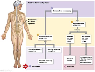
Additional info: The afferent (sensory) pathways transmit signals toward the CNS, while efferent (motor) pathways transmit signals away from the CNS to effectors.
Neural Tissue and Neuron Structure
Components of a Neuron
Neurons are the basic functional units of the nervous system, specialized for communication. Each neuron consists of several key structures:
Cell Body (Soma): Contains the nucleus and organelles; responsible for metabolic activities.
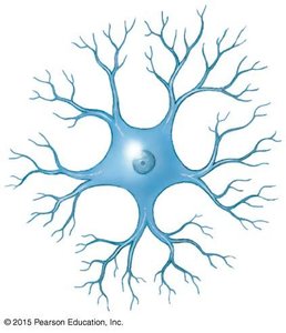
Dendrites: Highly branched extensions that receive incoming signals and convey them toward the cell body.
Axon: A single, long process that transmits electrical impulses away from the cell body to other neurons or effectors.
Axon Hillock: The origin of the axon from the cell body; site where action potentials are initiated.
Initial Segment: The first part of the axon where action potentials begin.
Telodendria: Fine extensions at the end of the axon.
Axon Terminals: The ends of telodendria where communication with other cells occurs.
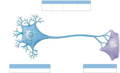
Additional info: The perikaryon is the cytoplasm surrounding the nucleus, containing organelles that provide energy and synthesize neurotransmitters.
Types of Synapses
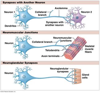
Neurons communicate with other cells at specialized junctions called synapses. There are three main types:
Synapses with Another Neuron: The axon terminal of one neuron communicates with the dendrite or cell body of another neuron.
Neuromuscular Junctions: The axon terminal communicates with a skeletal muscle fiber.
Neuroglandular Synapses: The axon terminal communicates with a gland cell.
Classification of Neurons
Structural Classification
Neurons are classified based on the number of processes extending from the cell body:
Anaxonic Neurons: Have no obvious axon; found in the brain; function is not well understood.
Unipolar Neurons: Have a single process that branches into dendrite and axon; common in sensory neurons of the PNS.
Bipolar Neurons: Have two distinct processes (one dendrite, one axon); found in special sensory organs.
Multipolar Neurons: Have one axon and two or more dendrites; most common type in the CNS and all motor neurons controlling skeletal muscles.
Functional Classification
Neurons are also classified by function:
Sensory Neurons: Transmit impulses from sensory receptors toward the CNS; mostly unipolar.
Motor Neurons: Carry impulses from the CNS to effectors; multipolar.
Interneurons: Lie between sensory and motor neurons; responsible for higher functions such as memory, learning, and planning; make up 99% of all neurons.
Neuroglia (Glial Cells)
Types and Functions
Neuroglia are supporting cells that protect and assist neurons. They are abundant and diverse, making up about half the volume of the nervous system.
CNS Neuroglia:
Astrocytes: Form the blood-brain barrier, isolating the CNS from chemicals and hormones in the blood.
Microglia: Remove waste and pathogens; related to monocytes and macrophages.
Ependymal Cells: Line CNS passageways filled with cerebrospinal fluid.
Oligodendrocytes: Provide structural framework and form myelin sheaths in the CNS, increasing the speed of nerve impulse transmission.
PNS Neuroglia:
Schwann Cells: Cover peripheral axons, participate in repair after injury, and form myelin sheaths in the PNS.
Satellite Cells: Cover neuron cell bodies in the PNS, functioning similarly to astrocytes.
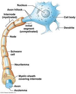
Additional info: Myelin sheaths are segmented coverings that protect and electrically insulate axons, allowing for faster transmission of nerve impulses. Multiple sclerosis is a disease where myelin is destroyed.
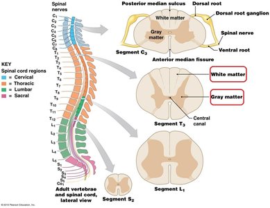
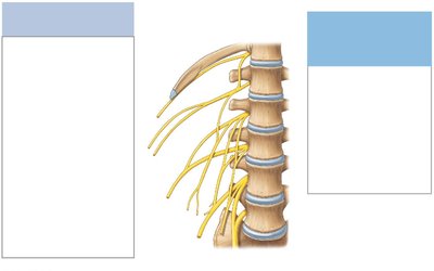
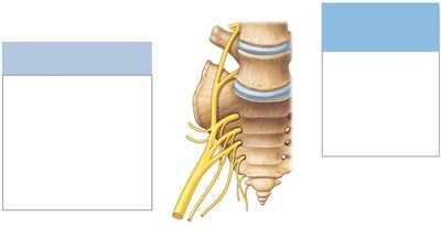
Spinal Cord Structure and Organization
Regions and Enlargements of the Spinal Cord
The spinal cord is divided into regions and contains enlargements where nerves serving the limbs arise.
Cervical Enlargement: Innervates the shoulders and upper limbs.
Lumbar Enlargement: Innervates the pelvis and lower limbs.
Conus Medullaris: Tapered, cone-shaped end of the spinal cord.
Cauda Equina: Bundle of nerve roots extending below the spinal cord.
Filum Terminale: Fibrous tissue extending from the conus medullaris to the coccyx, providing support.
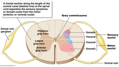
Cross-Sectional Anatomy of the Spinal Cord
The spinal cord consists of white and gray matter, roots, and nerves:
White Matter: Contains myelinated axons; transmits signals up and down the cord.
Gray Matter: Contains neuron cell bodies, dendrites, and unmyelinated axons; shaped like a butterfly.
Dorsal (Posterior) Root: Contains sensory neuron axons.
Ventral (Anterior) Root: Contains motor neuron axons.
Spinal Nerve: Contains both sensory and motor axons.
Functional Organization of Gray Matter
Gray matter in the spinal cord is organized into horns and commissures:
Posterior Gray Horn: Contains somatic and visceral sensory nuclei.
Anterior Gray Horn: Contains somatic motor nuclei.
Lateral Gray Horn: Contains visceral motor nuclei (in thoracic and upper lumbar regions).
Gray Commissures: Axons crossing from one side of the cord to the other.
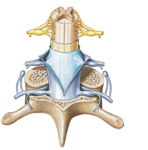
Spinal Nerves and Plexuses
Connective Tissue Layers of a Spinal Nerve
Each spinal nerve is surrounded by three connective tissue layers:
Epineurium: Outermost layer; surrounds the entire nerve.
Perineurium: Surrounds bundles of axons (fascicles).
Endoneurium: Surrounds individual axons.
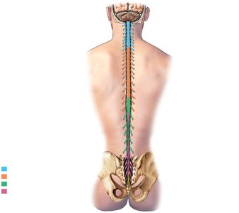
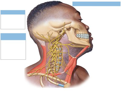
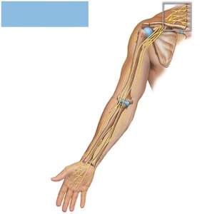
Major Nerve Plexuses
Plexuses are networks of intersecting nerves. The major plexuses include:
Cervical Plexus: Innervates the neck, back of the head, and diaphragm.
Brachial Plexus: Innervates the pectoral girdle and upper limbs.
Lumbar Plexus: Innervates the pelvic girdle and lower limbs.
Sacral Plexus: Innervates the pelvic region and lower limbs.
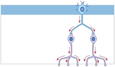
Neuronal Circuit Patterns
Types of Circuit Patterns
Neuronal circuits determine how information is processed in the nervous system:
Divergence: One neuron sends information to multiple neurons, allowing broad distribution of a signal.
Convergence: Multiple neurons synapse on a single postsynaptic neuron, integrating information from several sources.
Serial Processing: Information is relayed in a stepwise fashion from one neuron to another.
Parallel Processing: Several neurons process the same information simultaneously.
Reverberation: Collateral branches of axons extend back toward the source, creating a feedback loop.
Reflexes and Reflex Arcs
Steps in a Reflex Arc
Reflexes are rapid, automatic responses to stimuli. The basic steps in a reflex arc are:
Arrival of stimulus and activation of receptor
Activation of sensory neuron
Information processing in the CNS
Activation of a motor neuron
Response by effector
Additional info: Reflexes can be classified as innate or acquired, somatic or visceral, monosynaptic or polysynaptic, and spinal or cranial based on their development, response, complexity, and processing site.
