 Back
BackThe Nervous System: Structure, Function, and Anatomy
Study Guide - Smart Notes
 Tailored notes based on your materials, expanded with key definitions, examples, and context.
The Nervous System
Structural and Functional Classification
The nervous system is divided into structural and functional categories to better understand its organization and roles in the human body.
Structural Classification:
Central Nervous System (CNS): Consists of the brain and spinal cord. Responsible for integrating, processing, and coordinating sensory data and motor commands.
Peripheral Nervous System (PNS): Includes all nerves outside the CNS. Connects the CNS to limbs and organs.
Functional Classification:
Sensory (Afferent) Division: Transmits sensory information from receptors to the CNS.
Motor (Efferent) Division: Carries commands from the CNS to effectors (muscles and glands).
Somatic Nervous System: Controls voluntary movements of skeletal muscles.
Autonomic Nervous System: Regulates involuntary functions (e.g., heart rate, digestion). Subdivided into sympathetic and parasympathetic systems.
Neuron Anatomy and Types of Cells in the Nervous System
Neuron Structure
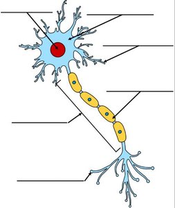
Neurons are specialized cells that transmit electrical impulses. Their anatomy is essential for understanding nerve function.
Dendrites: Receive signals from other neurons.
Cell Body (Soma): Contains the nucleus and organelles; integrates incoming signals.
Axon: Conducts electrical impulses away from the cell body.
Myelin Sheath: Insulates the axon, speeding up impulse transmission.
Nodes of Ranvier: Gaps in the myelin sheath where action potentials are regenerated.
Axon Terminals: Release neurotransmitters to communicate with other cells.
Types of Cells in the Nervous System
Neurons: Primary signaling cells.
Glial Cells: Support and protect neurons. Types include:
Astrocytes: Maintain blood-brain barrier, provide nutrients.
Oligodendrocytes: Form myelin in CNS.
Schwann Cells: Form myelin in PNS.
Microglia: Immune defense in CNS.
Ependymal Cells: Line ventricles, produce cerebrospinal fluid.
Nerve Transmission: Action Potential
Mechanism of Action Potential
An action potential is a rapid change in membrane potential that allows neurons to transmit signals.
Resting Potential: The neuron membrane is polarized, with a typical value of -70 mV.
Depolarization: Sodium channels open, Na+ enters, membrane potential becomes positive.
Repolarization: Potassium channels open, K+ exits, membrane potential returns to negative.
Hyperpolarization: Membrane potential temporarily becomes more negative than resting.
Propagation: Action potential travels along the axon, jumping between nodes of Ranvier.
Equation:
Example: The transmission of a pain signal from a finger to the brain involves action potentials traveling along sensory neurons.
CNS Structures
Major Structures of the Brain
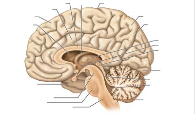
The central nervous system includes the brain, which is divided into several regions, each with distinct functions.
Cerebrum: Responsible for higher brain functions such as thought, memory, and voluntary movement.
Cerebellum: Coordinates movement and balance.
Brainstem: Controls basic life functions (breathing, heart rate).
Thalamus: Relay station for sensory information.
Hypothalamus: Regulates homeostasis, hormone release.
Corpus Callosum: Connects left and right hemispheres.
Pituitary Gland: Master endocrine gland.
PNS: Cranial Nerves, Spinal Nerves, and Plexuses
Cranial Nerves
Cranial nerves emerge directly from the brain and are responsible for sensory and motor functions of the head and neck.
12 pairs of cranial nerves: Each has specific functions (e.g., olfactory for smell, optic for vision).
Classification: Sensory, motor, or mixed.
Spinal Nerves and Plexuses
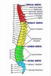
Spinal nerves arise from the spinal cord and are grouped into plexuses that serve different body regions.
Cervical Plexus: Serves head, neck, diaphragm.
Brachial Plexus: Serves arms.
Lumbar Plexus: Serves legs.
Sacral Plexus: Serves pelvic region.
Sympathetic and Parasympathetic Systems
Autonomic Nervous System Divisions
The autonomic nervous system is divided into sympathetic and parasympathetic branches, each with distinct effects on the body.
Sympathetic System: Prepares the body for 'fight or flight' responses. Increases heart rate, dilates pupils, inhibits digestion.
Parasympathetic System: Promotes 'rest and digest' activities. Decreases heart rate, constricts pupils, stimulates digestion.
Example: During stress, the sympathetic system increases heart rate; after eating, the parasympathetic system stimulates digestion.
Cranial Nerves: Names and Functions
Overview of Cranial Nerves
There are twelve cranial nerves, each with specific sensory, motor, or mixed functions.
I. Olfactory: Sensory - smell
II. Optic: Sensory - vision
III. Oculomotor: Motor - eye movement
IV. Trochlear: Motor - eye movement
V. Trigeminal: Mixed - facial sensation, chewing
VI. Abducens: Motor - eye movement
VII. Facial: Mixed - facial expression, taste
VIII. Vestibulocochlear: Sensory - hearing, balance
IX. Glossopharyngeal: Mixed - taste, swallowing
X. Vagus: Mixed - autonomic control of heart, lungs, digestive tract
XI. Accessory: Motor - neck muscles
XII. Hypoglossal: Motor - tongue movement
Classification Table:
Nerve | Type | Main Function |
|---|---|---|
Olfactory (I) | Sensory | Smell |
Optic (II) | Sensory | Vision |
Oculomotor (III) | Motor | Eye movement |
Trigeminal (V) | Mixed | Facial sensation, chewing |
Facial (VII) | Mixed | Facial expression, taste |
Vagus (X) | Mixed | Autonomic control |
