 Back
BackThe Peripheral Nervous System: Structure, Function, and Organization
Study Guide - Smart Notes
 Tailored notes based on your materials, expanded with key definitions, examples, and context.
The Peripheral Nervous System (PNS)
Overview of the PNS
The Peripheral Nervous System (PNS) consists of all neural structures outside the brain and spinal cord. It includes sensory receptors, peripheral nerves, associated ganglia, and motor endings. The PNS provides essential communication links between the body and the central nervous system (CNS), allowing the body to respond to internal and external stimuli.
Sensory receptors detect changes in the environment and initiate nerve impulses.
Peripheral nerves transmit these impulses to and from the CNS.
Ganglia are clusters of neuron cell bodies in the PNS.
Motor endings activate effector organs such as muscles and glands.
Sensory Receptors
Classification by Stimulus Type
Sensory receptors are specialized to respond to specific types of stimuli. Their activation leads to depolarization and the generation of nerve impulses, which are interpreted by the brain as sensations and perceptions.
Mechanoreceptors: Respond to mechanical forces such as touch, pressure, vibration, stretch, and itch.
Thermoreceptors: Detect changes in temperature.
Photoreceptors: Respond to light energy (e.g., in the retina).
Chemoreceptors: Respond to chemical stimuli (e.g., smell, taste, changes in blood chemistry).
Nociceptors: Detect potentially damaging stimuli that result in pain.
Classification by Location
Exteroceptors: Located near the body surface; respond to external stimuli such as touch, pressure, pain, and temperature. Include special sense organs.
Interoceptors (Visceroceptors): Located in internal organs and blood vessels; respond to internal stimuli such as chemical changes, stretch, and temperature.
Proprioceptors: Found in muscles, tendons, joints, and connective tissues; monitor the degree of stretch and position of body parts, providing information about body movement.
Classification by Structural Complexity
Sensory receptors are also classified as simple or complex based on their structure:
Simple receptors: Most common; include encapsulated and unencapsulated varieties. Detect general sensations such as touch, pain, and temperature.
Complex receptors: Special sense organs (e.g., eyes, ears, nose, taste buds).
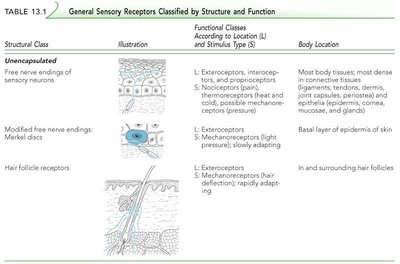
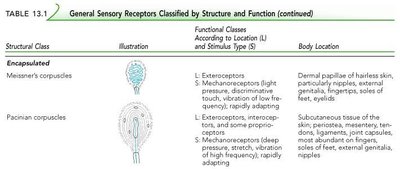
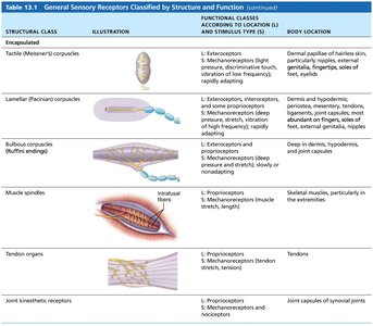
Simple Receptors: Unencapsulated and Encapsulated
Unencapsulated receptors are free nerve endings, while encapsulated receptors are enclosed in connective tissue capsules. Both types are distributed throughout the body and are responsible for detecting a variety of sensory modalities.
Unencapsulated: Free nerve endings, Merkel (tactile) discs, hair follicle receptors.
Encapsulated: Meissner's corpuscles, Pacinian corpuscles, Ruffini endings, muscle spindles, Golgi tendon organs, joint kinesthetic receptors.
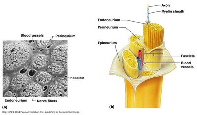
Structure of a Nerve
Organization and Connective Tissue Coverings
A nerve is a cordlike organ of the PNS, consisting of bundles of peripheral axons (nerve fibers) enclosed by connective tissue. The connective tissue layers are:
Endoneurium: Surrounds individual axons.
Perineurium: Bundles groups of axons into fascicles.
Epineurium: Encloses all fascicles to form the nerve.
Classification of Nerves
Sensory, Motor, and Mixed Nerves
Nerves are classified based on the direction of impulse transmission:
Sensory (afferent) nerves: Carry impulses toward the CNS.
Motor (efferent) nerves: Carry impulses away from the CNS.
Mixed nerves: Contain both sensory and motor fibers; most common type in the body.
Mixed nerves may carry somatic (voluntary) and autonomic (involuntary) fibers, including somatic afferent/efferent and visceral afferent/efferent fibers.
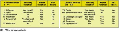
Cranial Nerves
Overview and Classification
There are twelve pairs of cranial nerves that arise from the brain. Each is identified by a Roman numeral (I–XII) and a name. Cranial nerves may be sensory, motor, or mixed, and some carry parasympathetic fibers.
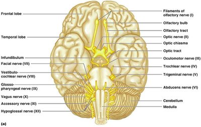
Individual Cranial Nerves
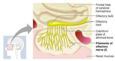
I. Olfactory: Sensory for smell; fibers pass through the cribriform plate to the olfactory cortex.
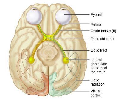
II. Optic: Sensory for vision; fibers arise from the retina, pass through the optic chiasm, and project to the visual cortex.
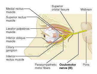
III. Oculomotor: Motor to most extrinsic eye muscles; controls eyelid, iris, and lens shape.
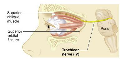
IV. Trochlear: Motor to the superior oblique muscle of the eye.
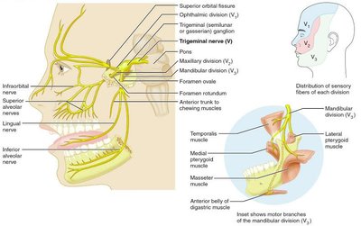
V. Trigeminal: Mixed; sensory from face, motor to muscles of mastication. Three divisions: ophthalmic (V1), maxillary (V2), mandibular (V3).
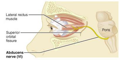
VI. Abducens: Motor to the lateral rectus muscle of the eye.
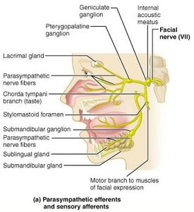
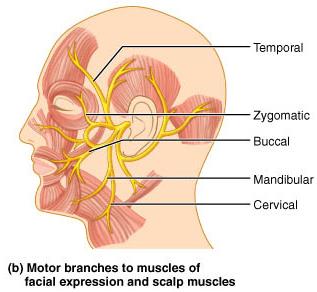
VII. Facial: Mixed; motor to muscles of facial expression, parasympathetic to glands, sensory for taste (anterior 2/3 of tongue).
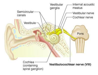
VIII. Vestibulocochlear: Sensory for hearing and equilibrium; cochlear and vestibular divisions.
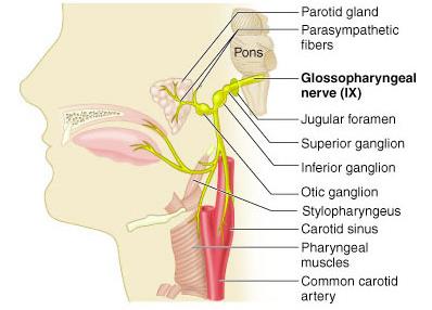
IX. Glossopharyngeal: Mixed; motor to pharynx and parotid gland, sensory for taste and general sensation from tongue and pharynx.
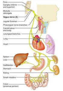
X. Vagus: Mixed; only cranial nerve to extend beyond head and neck, parasympathetic to thoracic and abdominal organs, sensory for taste.
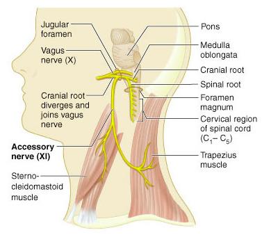
XI. Accessory: Motor to sternocleidomastoid and trapezius muscles; cranial and spinal roots.
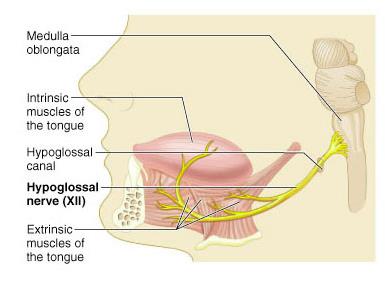
XII. Hypoglossal: Motor to intrinsic and extrinsic muscles of the tongue.
Spinal Nerves and Plexuses
Spinal Nerves
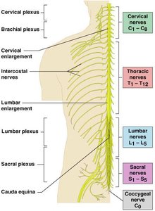
There are 31 pairs of spinal nerves, all of which are mixed nerves. They are named according to their region of origin:
8 cervical (C1–C8)
12 thoracic (T1–T12)
5 lumbar (L1–L5)
5 sacral (S1–S5)
1 coccygeal (C0)
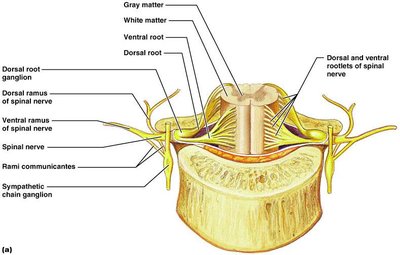
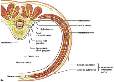
Roots and Rami of Spinal Nerves
Each spinal nerve connects to the spinal cord via two roots:
Ventral root: Contains motor (efferent) fibers from the anterior horn.
Dorsal root: Contains sensory (afferent) fibers from the dorsal root ganglion.
After emerging from the spinal cord, each spinal nerve branches into rami:
Dorsal ramus: Innervates the back.
Ventral ramus: Innervates the limbs and anterior body wall.
Meningeal branch: Reenters the vertebral canal to innervate meninges and blood vessels.
Rami communicantes: Contain autonomic fibers (in thoracic region).
Nerve Plexuses
Ventral rami (except T2–T12) form interlacing networks called plexuses in the cervical, brachial, lumbar, and sacral regions. Each plexus gives rise to nerves that serve specific body regions.
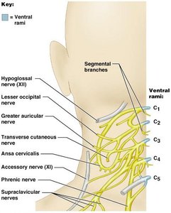
Cervical plexus (C1–C4): Innervates neck, ear, back of head, and shoulders; phrenic nerve supplies the diaphragm.
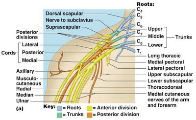
Brachial plexus (C5–T1): Innervates upper limb; major nerves include axillary, musculocutaneous, median, radial, and ulnar.
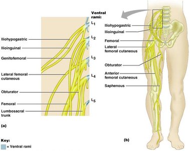
Lumbar plexus (L1–L4): Innervates thigh, abdominal wall, and psoas muscle; major nerves are femoral, obturator, and saphenous.
Sacral plexus (L4–S4): Innervates buttock, lower limb, pelvic structures, and perineum; major nerve is the sciatic (tibial and common fibular nerves).
Dermatomes
A dermatome is an area of skin innervated by the cutaneous branches of a single spinal nerve. All spinal nerves except C1 participate in dermatomes, which are important for diagnosing nerve injuries.
Innervation of Joints
According to Hilton’s law, any nerve serving a muscle that produces movement at a joint also innervates the joint itself and the skin over the joint. This ensures coordinated sensory and motor function at joints.
