 Back
BackThe Reproductive System: Structure, Function, and Regulation
Study Guide - Smart Notes
 Tailored notes based on your materials, expanded with key definitions, examples, and context.
Introduction to the Male and Female Reproductive Systems
Overview
The reproductive systems in males and females are responsible for producing gametes, secreting sex hormones, and supporting the processes of fertilization and development. The primary sex organs, or gonads, are the testes in males and ovaries in females. These organs produce gametes (sperm and ova) and secrete hormones such as testosterone and estrogens. Accessory reproductive organs aid in the transport, nourishment, and protection of gametes.
Gonads: Primary sex organs; testes (males), ovaries (females).
Sex Hormones: Testosterone (males), estrogens (females).
Gametes: Sperm (males), ova (females); produced via meiosis.
Accessory Organs: Ducts, glands, and external genitalia that support reproductive function.
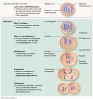
Meiosis and Gametogenesis
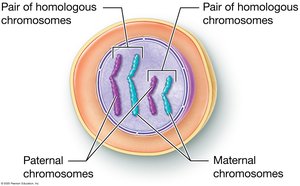
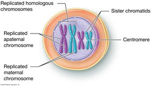
Chromosomes and Genetic Variation
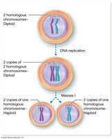
Somatic cells are diploid (2n), containing 46 chromosomes (23 pairs), with one set from each parent. Homologous chromosomes carry the same genes but may have different alleles. Gametes are haploid (1n), containing 23 chromosomes, ensuring the zygote formed at fertilization has the correct chromosome number.
Homologous Chromosomes: Chromosome pairs with the same genes.
Alleles: Variants of a gene found on homologous chromosomes.
Fertilization: Fusion of sperm and ovum to form a zygote.
Meiosis: Process and Phases
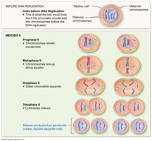
Meiosis is a specialized cell division that reduces chromosome number by half, producing four genetically unique haploid cells. It consists of two consecutive divisions: Meiosis I (reduction division) and Meiosis II (equational division).
Meiosis I: Homologous chromosomes separate, resulting in haploid cells.
Meiosis II: Sister chromatids separate, producing four unique gametes.
Key Events: DNA replication, synapsis, crossing over, independent assortment.

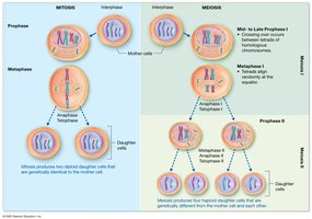
Comparison of Mitosis and Meiosis
Mitosis produces two genetically identical diploid cells for growth and repair, while meiosis produces four genetically unique haploid cells for sexual reproduction.
Mitosis: One division, no crossing over, identical cells.
Meiosis: Two divisions, crossing over, genetic diversity.
Anatomy of the Male Reproductive System
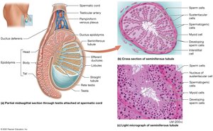
Testes
The testes are paired organs located in the scrotum, responsible for sperm production and hormone secretion. Seminiferous tubules within the testes contain spermatogenic and sustentacular cells, while interstitial cells produce testosterone.
Seminiferous Tubules: Site of spermatogenesis.
Interstitial Cells: Produce testosterone.
Myoid Cells: Aid in sperm transport.
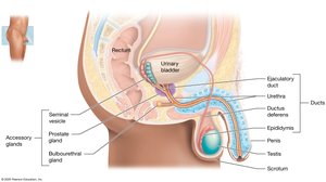
Duct System
Sperm travel from the testes through the epididymis, ductus deferens, ejaculatory duct, and urethra. Each region has specialized functions in sperm maturation, storage, and transport.
Epididymis: Sperm maturation and storage.
Ductus Deferens: Transports sperm during ejaculation.
Ejaculatory Duct: Joins ductus deferens and seminal vesicle duct.
Urethra: Passage for urine and semen.
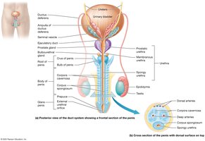
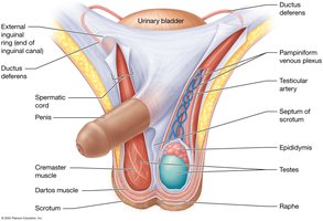
Penis and Scrotum
The penis is the male copulatory organ, containing erectile tissue that enables erection. The scrotum houses the testes and regulates their temperature for optimal sperm production.
Corpora Cavernosa and Corpus Spongiosum: Erectile tissues.
Prepuce (Foreskin): May be removed by circumcision.
Scrotum: Temperature regulation via cremaster and dartos muscles.
Accessory Sex Glands
Accessory glands produce seminal fluid, which nourishes and protects sperm. These include the seminal vesicles, prostate gland, and bulbourethral glands.
Seminal Vesicles: Secrete fructose-rich fluid (energy for sperm).
Prostate Gland: Secretes citrate, PSA, and antimicrobial chemicals.
Bulbourethral Glands: Secrete alkaline mucus for lubrication.
Spermatogenesis and Hormonal Regulation
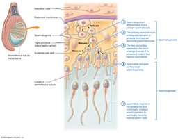
Spermatogenesis
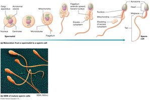
Spermatogenesis is the process of sperm cell development, beginning at puberty and continuing throughout life. It occurs in the seminiferous tubules and involves mitosis, meiosis, and spermiogenesis (maturation of spermatids into spermatozoa).
Spermatogonia: Diploid stem cells.
Primary Spermatocyte: Undergoes meiosis I.
Secondary Spermatocyte: Undergoes meiosis II.
Spermatids: Differentiate into mature sperm.
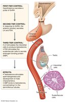
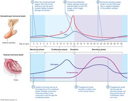
Hormonal Control (HPG Axis)
The hypothalamic-pituitary-gonadal (HPG) axis regulates male reproductive function. GnRH from the hypothalamus stimulates the anterior pituitary to release LH and FSH, which act on the testes to promote testosterone production and spermatogenesis.
GnRH: Stimulates LH and FSH release.
LH: Stimulates testosterone production by interstitial cells.
FSH: Stimulates sustentacular cells to support spermatogenesis.
Inhibin: Inhibits FSH secretion.
Anatomy of the Female Reproductive System
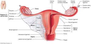
Ovaries
The ovaries are paired organs that produce oocytes and secrete hormones such as estrogens and progesterone. Oogenesis occurs in the ovarian cortex, where follicles at various stages of development are found.
Follicles: Structures containing developing oocytes.
Hormones: Estrogens, progesterone, inhibin, relaxin.
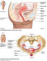
Uterine Tubes, Uterus, and Vagina
The uterine tubes (fallopian tubes) transport oocytes from the ovaries to the uterus, where fertilization may occur. The uterus supports implantation and development of the conceptus. The vagina serves as the organ of copulation and birth canal.
Fimbriae: Finger-like projections that capture oocytes.
Uterus: Site of implantation, fetal development, and menstruation.
Vagina: Receives penis and semen; passageway for childbirth and menstruation.
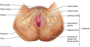
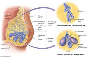
External Genitalia and Mammary Glands
The vulva includes the mons pubis, labia majora and minora, clitoris, and vestibular glands. Mammary glands are modified sweat glands that produce milk for newborns.
Clitoris: Erectile tissue important in sexual response.
Mammary Glands: Produce and secrete milk.
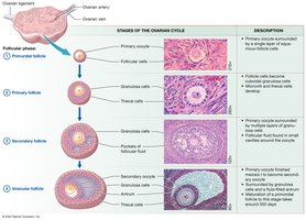
Oogenesis and the Ovarian Cycle
Oogenesis
Oogenesis is the process of ovum development, beginning before birth and continuing until menopause. It involves mitosis, meiosis, and the formation of polar bodies to conserve cytoplasm for the ovum.
Oogonia: Stem cells divide by mitosis before birth.
Primary Oocytes: Arrested in prophase I until puberty.
Secondary Oocyte: Released at ovulation; completes meiosis II only if fertilized.
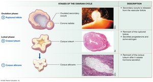
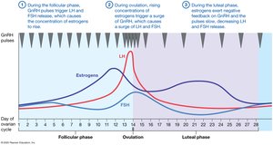
Ovarian Cycle
The ovarian cycle consists of the follicular phase (follicle growth), ovulation (release of secondary oocyte), and luteal phase (formation of corpus luteum). Hormonal regulation involves GnRH, LH, FSH, estrogens, and progesterone.
Follicular Phase: Follicle maturation and estrogen production.
Ovulation: LH surge triggers release of secondary oocyte.
Luteal Phase: Corpus luteum secretes progesterone; degenerates if no pregnancy.
The Uterine (Menstrual) Cycle
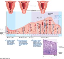
Phases of the Uterine Cycle
The uterine cycle describes changes in the endometrium in response to ovarian hormones. It includes the menstrual phase (shedding of the functional layer), proliferative phase (regeneration), and secretory phase (preparation for implantation).
Menstrual Phase (Days 1–5): Shedding of stratum functionalis.
Proliferative Phase (Days 6–14): Endometrial regeneration.
Secretory Phase (Days 15–28): Glandular secretion and preparation for implantation.
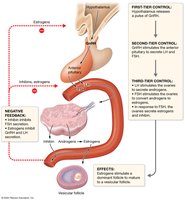
Hormonal Regulation of Reproduction
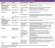
Major Reproductive Hormones
Reproductive function is regulated by a complex interplay of hormones from the hypothalamus, pituitary gland, and gonads. These hormones control gametogenesis, secondary sex characteristics, and reproductive cycles.
Hormone | Source | Target Tissue(s) | Response |
|---|---|---|---|
GnRH | Hypothalamus | Anterior pituitary | Stimulates secretion of LH and FSH |
LH | Anterior pituitary | Testes, ovaries | Stimulates testosterone/estrogen production, ovulation |
FSH | Anterior pituitary | Testes, ovaries | Stimulates gamete production |
Testosterone | Testes | Various | Male secondary sex characteristics, spermatogenesis |
Estrogens | Ovaries | Various | Female secondary sex characteristics, endometrial growth |
Progesterone | Ovaries | Uterus, mammary glands | Prepares uterus for implantation, maintains pregnancy |
Inhibin | Testes, ovaries | Anterior pituitary | Inhibits FSH secretion |

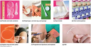
Birth Control Methods
Overview of Contraceptive Methods
Birth control methods are designed to prevent pregnancy by various mechanisms, including behavioral, barrier, hormonal, intrauterine, and permanent (sterilization) methods. Effectiveness varies by method and user compliance.
Behavioral: Abstinence, rhythm method, withdrawal.
Barrier: Condoms, diaphragms, cervical caps, sponges.
Hormonal: Oral contraceptives, injections, implants, patches, rings.
Intrauterine: IUDs, IUSs.
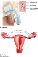
Permanant: Vasectomy, tubal ligation, tubal implants.
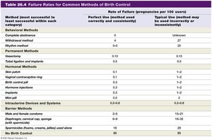
Method | Perfect Use Failure Rate (%) | Typical Use Failure Rate (%) |
|---|---|---|
Abstinence | 0 | Unknown |
Male Condom | 2 | 18 |
Oral Contraceptives | 0.3 | 9 |
IUD | 0.6 | 0.8 |
Vasectomy | 0.1 | 0.15 |
No Birth Control | 85 | 85 |
Other methods | See table | See table |
Sexually Transmitted Infections (STIs)
Overview and Common STIs
STIs are infections transmitted through sexual contact and are a major cause of reproductive disorders and infertility. They may be caused by bacteria, viruses, or parasites.
Bacterial: Chlamydia, gonorrhea, syphilis.
Parasitic: Trichomoniasis.
Viral: HPV (genital warts), genital herpes.
Cervical Cancer and HPV
Cervical cancer is often linked to HPV infection and is most common in women aged 30–50. Early detection via Pap smears and HPV vaccination are key preventive measures.

Summary Table: Functions of Major Reproductive Structures
Structure | Function |
|---|---|
Testes | Produce sperm and testosterone |
Epididymis | Sperm maturation and storage |
Ductus Deferens | Transports sperm during ejaculation |
Seminal Vesicles | Produce seminal fluid |
Prostate Gland | Secretes prostatic fluid |
Ovaries | Produce oocytes and hormones |
Uterine Tubes | Transport oocyte; site of fertilization |
Uterus | Supports fetal development |
Vagina | Receives penis; birth canal |
Additional info: This guide integrates foundational concepts of reproductive anatomy and physiology, gametogenesis, hormonal regulation, and clinical relevance, providing a comprehensive overview for college-level study.
