 Back
BackThe Reproductive System: Structure, Function, and Cellular Processes
Study Guide - Smart Notes
 Tailored notes based on your materials, expanded with key definitions, examples, and context.
The Reproductive System
Overview of the Reproductive System
The reproductive system is responsible for the production of gametes, secretion of sex hormones, and the facilitation of fertilization. It consists of primary sex organs (gonads), accessory reproductive organs, and external genitalia. Gonads produce gametes—sperm in males and ova in females—and secrete sex hormones such as androgens (testosterone) in males and estrogens/progesterone in females.
Primary Sex Organs: Testes in males, ovaries in females
Accessory Organs: Ducts, glands, and external genitalia
Sex Hormones: Androgens (testosterone), estrogens, progesterone
Cell Division: Mitosis vs. Meiosis
Comparison of Mitosis and Meiosis
Mitosis and meiosis are two distinct processes of cell division. Mitosis results in two genetically identical diploid cells, while meiosis produces four genetically unique haploid gametes. Meiosis involves two consecutive divisions: Meiosis I (reductional division) and Meiosis II (equational division).
Mitosis: One division, produces diploid cells (2n), identical to parent
Meiosis: Two divisions, produces haploid cells (n), genetically unique
Meiosis I: Homologous chromosomes separate, chromosome number halved
Meiosis II: Sister chromatids separate, maintains haploid number
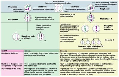
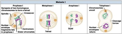
Meiosis I: Reductional Division
Meiosis I reduces the chromosome number from diploid (2n) to haploid (n). It includes prophase I (synapsis and crossing over), metaphase I (tetrads align), anaphase I (homologous chromosomes separate), and telophase I (chromosomes uncoil).
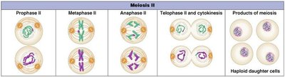
Meiosis II: Equational Division
Meiosis II resembles mitosis, where sister chromatids separate, resulting in four haploid daughter cells. In males, this produces four sperm; in females, one ovum and three polar bodies.
Male Reproductive System
Anatomy and Function
The male reproductive system includes the testes, ducts, accessory glands, and penis. The testes produce sperm and testosterone. Sperm travel through the epididymis, ductus deferens, ejaculatory duct, and urethra. Accessory glands (seminal vesicles, prostate, bulbourethral glands) contribute to semen production.
Testes: Sperm production, testosterone secretion
Ducts: Epididymis, ductus deferens, ejaculatory duct, urethra
Accessory Glands: Seminal vesicles, prostate, bulbourethral glands
Penis: Erectile tissue, urethra for sperm delivery
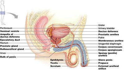
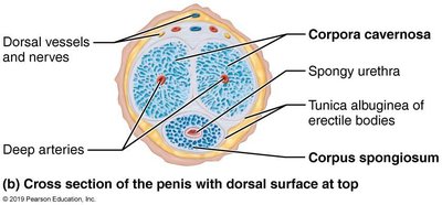
Penis Structure
The penis contains three cylindrical bodies of erectile tissue: corpus spongiosum (surrounds urethra), and paired corpora cavernosa (dorsal). The tunica albuginea binds the corpora cavernosa.
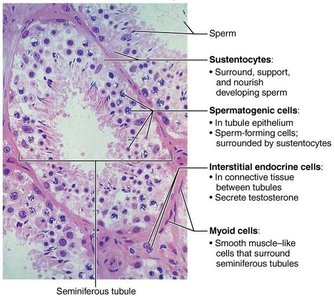
Testes and Seminiferous Tubules
The testes are divided into lobules containing seminiferous tubules, the site of sperm production. Interstitial cells between tubules produce testosterone.
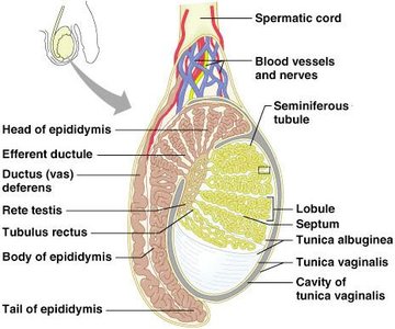
Epididymis
The epididymis stores and matures sperm. Stereocilia absorb fluid and pass nutrients to sperm. Upon ejaculation, sperm are expelled into the ductus deferens.
Ductus Deferens and Ejaculatory Duct
The ductus deferens transports sperm from the epididymis to the urethra. Its terminus forms the ampulla and joins the seminal vesicle duct to form the ejaculatory duct. Vasectomy involves cutting the ductus deferens for birth control.
Accessory Glands
Seminal Vesicles: Secrete alkaline fluid with fructose, prostaglandins, and enzymes
Prostate Gland: Produces milky, acidic fluid with citrate, enzymes, and PSA
Bulbourethral Glands: Secrete alkaline mucus to neutralize urine acidity
Semen
Semen is a mixture of sperm and glandular secretions. It provides nutrients, protects sperm, and facilitates movement. Prostaglandins decrease cervical mucus viscosity and stimulate uterine contractions. Relaxin enhances sperm motility.
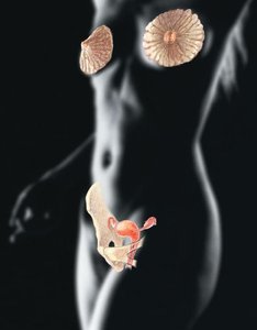
Female Reproductive System
Anatomy and Function
The female reproductive system includes the ovaries, uterine tubes, uterus, vagina, and external genitalia. Ovaries produce ova and secrete estrogen and progesterone. Accessory ducts transport gametes and facilitate fertilization.
Ovaries: Primary female reproductive organs
Uterine Tubes: Site of fertilization, transport oocyte
Uterus: Site of implantation and development
Vagina: Birth canal, copulation organ
External Genitalia: Mons pubis, labia, clitoris
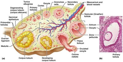
Ovarian Structure
Ovaries are surrounded by tunica albuginea and germinal epithelium. The cortex contains follicles at various stages of development. Each follicle consists of an oocyte surrounded by follicular or granulosa cells.

Follicle Development
Primordial Follicle: One layer of squamous follicle cells
Primary Follicle: Multiple layers of cuboidal granulosa cells
Secondary Follicle: Fluid-filled antrum forms
Graafian Follicle: Mature follicle ready for ovulation
Corpus Luteum: Formed after ovulation, secretes hormones
Uterine Tubes (Fallopian Tubes)
Uterine tubes receive the ovulated oocyte and provide a site for fertilization. The ampulla is the main site of fertilization, and fimbriae help capture the oocyte.
Uterus
The uterus is a hollow, muscular organ with three layers: perimetrium (outer), myometrium (muscle), and endometrium (mucosal lining). The endometrium has a functional layer (shed during menstruation) and a basal layer (regenerates functional layer).
Uterine Vascular Supply
Uterine arteries branch into arcuate and radial arteries, supplying the endometrium. Spiral arteries supply the functional layer and degenerate during menstruation.
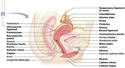
Vagina and External Genitalia
The vagina is a muscular tube for birth, menstrual flow, and copulation. The vulva includes the mons pubis, labia majora/minora, clitoris, and vestibular structures.
Mammary Glands
Mammary glands are modified sweat glands that produce milk. They consist of lobes, alveoli, and lactiferous ducts. Breast cancer often arises from ductal epithelial cells.
Gamete Formation: Spermatogenesis and Oogenesis
Spermatogenesis
Spermatogenesis is the process of sperm production in the seminiferous tubules. It involves mitosis of spermatogonia, meiosis of primary spermatocytes, and maturation of spermatids into sperm.
Spermatogonia: Stem cells undergo mitosis
Primary Spermatocytes: Undergo meiosis I
Secondary Spermatocytes: Undergo meiosis II
Spermatids: Mature into sperm (spermiogenesis)
Oogenesis
Oogenesis is the production of female gametes by meiosis. Oogonia divide by mitosis and become primary oocytes, which begin meiosis but stall in prophase I. At puberty, primary oocytes complete meiosis I, producing a secondary oocyte and a polar body. The secondary oocyte is ovulated and completes meiosis II if fertilized.
Hormonal Regulation
Gonadotropins and the HPG Axis
Gonadotropins (FSH and LH) regulate gamete production and hormone secretion. The hypothalamic-pituitary-gonadal (HPG) axis controls the release of GnRH, FSH, and LH, which act on the gonads.
FSH: Stimulates gamete production
LH: Stimulates hormone production and ovulation
Feedback: Rising testosterone and inhibin inhibit GnRH, FSH, and LH
Ovarian and Uterine Cycles
Ovarian Cycle
The ovarian cycle consists of the follicular phase (follicle growth), ovulation (release of oocyte), and luteal phase (corpus luteum activity). Hormonal interactions regulate these phases.
Follicular Phase: FSH and LH stimulate follicle growth; rising estrogen inhibits FSH/LH
Ovulation: LH surge triggers ovulation
Luteal Phase: Corpus luteum secretes progesterone and estrogen; if no pregnancy, degenerates
Uterine (Menstrual) Cycle
The uterine cycle involves cyclic changes in the endometrium in response to ovarian hormones. It includes the menstrual phase (shedding), proliferative phase (rebuilding), and secretory phase (preparation for implantation).
Menstrual Phase: Days 1-5, functional layer shed
Proliferative Phase: Days 6-14, endometrium rebuilds
Secretory Phase: Days 15-28, endometrium prepares for embryo
Summary Table: Mitosis vs. Meiosis
Event | Mitosis | Meiosis |
|---|---|---|
Number of divisions | One | Two |
Number of daughter cells | Two (diploid, identical) | Four (haploid, unique) |
Importance | Growth, repair, genetic stability | Gamete production, genetic diversity |
Additional info: All explanations have been expanded for academic completeness and clarity. Images included are directly relevant to the anatomical and cellular processes described.
