 Back
BackThe Reproductive System: Structure, Function, and Physiology
Study Guide - Smart Notes
 Tailored notes based on your materials, expanded with key definitions, examples, and context.
Introduction to the Reproductive System
Overview
The reproductive system ensures the continuity of life by producing offspring. It consists of primary sex organs (gonads) and accessory structures, with distinct male and female anatomical and physiological features. The system is also responsible for the production of gametes via meiosis and the secretion of sex hormones.
26.1: Overview of the Reproductive System and Meiosis
Male and Female Reproductive Systems: Structure and Function
Gonads: Testes in males and ovaries in females; produce gametes and secrete sex hormones (testosterone, estrogens).
Accessory Organs: Include ducts, glands, and external genitalia that support gamete transport and reproductive function.
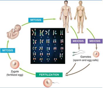
Human Sexual Life Cycle
Diploid (2n): Cells with two sets of chromosomes (all body cells except gametes).
Haploid (n): Gametes (sperm and ova) with one set of chromosomes, produced by meiosis.
Fertilization: Fusion of haploid gametes restores diploid chromosome number in the zygote.
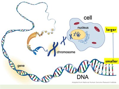
Cellular Basis of Reproduction
Chromosomes: Structures within the nucleus containing DNA and genes.
Gene: Segment of DNA coding for a specific protein.
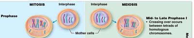
Mitosis vs. Meiosis
Mitosis: Cell division producing two genetically identical diploid cells; used for growth and repair.
Meiosis: Cell division producing four genetically unique haploid gametes; involves two successive divisions (Meiosis I and II).
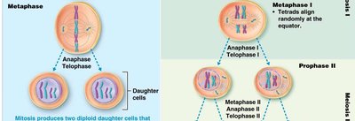
Key Differences Between Mitosis and Meiosis
Feature | Mitosis | Meiosis |
|---|---|---|
Number of Divisions | 1 | 2 |
Daughter Cells | 2 (diploid, identical) | 4 (haploid, unique) |
Function | Growth, repair | Gamete production |
26.2: Anatomy of the Male Reproductive System
Structure and Function
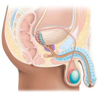
Testes: Produce sperm and testosterone; located in the scrotum.
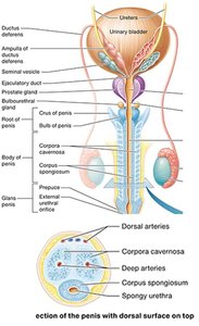
Ducts: Epididymis (sperm maturation), ductus deferens (sperm transport), ejaculatory ducts, and urethra (shared with urinary system).
Accessory Glands: Seminal vesicles, prostate gland, and bulbourethral glands add secretions to semen.
Penis: Organ of copulation; delivers sperm to the female reproductive tract.
Testes: Internal Anatomy and Histology
Tunica Vaginalis: Outer covering formed during testicular descent.
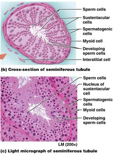
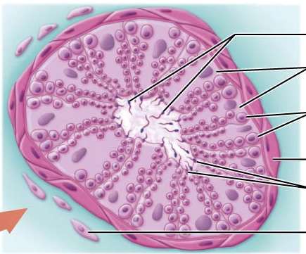
Tunica Albuginea: Dense connective tissue dividing testis into lobules (200-300 per testis), each containing seminiferous tubules (site of spermatogenesis).
Seminiferous Tubules: Lined with spermatogenic cells and sustentacular (Sertoli) cells; interstitial (Leydig) cells between tubules secrete testosterone.

Male Duct System and Penis
Epididymis: Sperm maturation and storage.
Ductus Deferens: Transports sperm during ejaculation.
Urethra: Prostatic, membranous, and spongy sections; conducts urine and semen.
Penis: Contains erectile tissues (corpora cavernosa and corpus spongiosum); glans penis covered by prepuce (foreskin).
Accessory Glands
Seminal Vesicles: Secrete alkaline fluid with fructose, prostaglandins, and clotting proteins.
Prostate Gland: Secretes citrate, PSA, and antimicrobial chemicals.
Bulbourethral Glands: Secrete mucus and alkaline fluid.
26.3: Physiology of the Male Reproductive System
Spermatogenesis
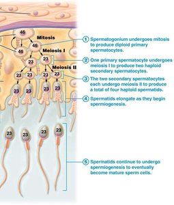
Spermatogonia: Diploid stem cells undergo mitosis to produce primary spermatocytes.
Meiosis I: Primary spermatocytes divide to form two haploid secondary spermatocytes.
Meiosis II: Each secondary spermatocyte divides to form two spermatids (total of four haploid cells).
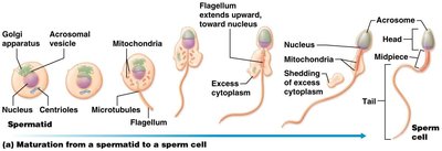
Spermiogenesis: Maturation of spermatids into spermatozoa.
Structure of Sperm
Head: Contains nucleus (23 chromosomes) and acrosomal cap (enzymes for oocyte penetration).
Midpiece: Packed with mitochondria for energy.
Tail: Flagellum for motility.
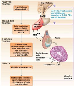
Hormonal Regulation
GnRH (Gonadotropin-releasing hormone): Released from hypothalamus; stimulates anterior pituitary to secrete LH and FSH.
FSH: Stimulates spermatogenesis via Sertoli cells.
LH: Stimulates Leydig cells to produce testosterone.
Testosterone: Promotes development of male secondary sex characteristics and spermatogenesis.
26.4: Anatomy of the Female Reproductive System
Structure and Function
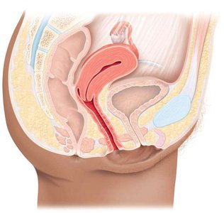
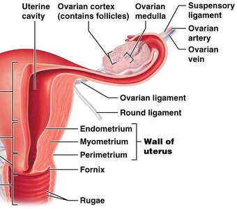
Ovaries: Produce ova and hormones; anchored by ligaments; cortex contains follicles, medulla contains blood vessels and nerves.
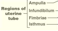
Uterine Tubes: Transport ova, sperm, and zygotes; regions include fimbriae, infundibulum, ampulla, and isthmus.
Uterus: Site of menstruation, implantation, fetal development, and labor; regions include fundus, body, isthmus, and cervix.
Vagina: Passage for sperm, menstrual flow, and childbirth.
External Genitalia (Vulva): Includes mons pubis, labia majora/minora, clitoris (homologous to male structures).
Mammary Glands: Modified sweat glands for milk production (lactation).
26.5: Physiology of the Female Reproductive System
Oogenesis
Oogenesis: Formation of haploid ova; begins before birth, pauses, resumes at puberty, and continues until menopause.
Folliculogenesis: Development of ovarian follicles; primordial, primary, secondary, and vesicular (Graafian) stages.
Ovulation: Release of secondary oocyte from vesicular follicle, triggered by LH surge.
Corpus Luteum: Secretes estrogen, progesterone, relaxin, and inhibin; degenerates to corpus albicans if no pregnancy occurs.
Female Reproductive (Menstrual) Cycle
Ovarian Cycle: Follicular phase (follicle development), ovulation, luteal phase (corpus luteum activity).
Uterine Cycle: Menstrual phase (shedding endometrium), proliferative phase (endometrial growth), secretory phase (preparation for implantation).
Hormonal Regulation: FSH and LH from anterior pituitary; estrogen and progesterone from ovaries.
Comparison: Spermatogenesis vs. Oogenesis
Feature | Spermatogenesis | Oogenesis |
|---|---|---|
Onset | Puberty | Before birth |
Process | Continuous | Cyclic |
Duration | Throughout life | Until menopause |
Gametes Produced | Millions/day | 1/month |
26.6: Methods of Birth Control
Barrier Methods: Prevent sperm from reaching oocyte (condoms, diaphragm).
Hormonal Methods: Inhibit ovulation (oral contraceptives, injections, vaginal rings).
Sterilization: Permanent prevention of gamete transport (vasectomy, tubal ligation).
IUDs: Prevent implantation of zygote.
Chemical Methods: Spermicides destroy sperm.
Behavioral Methods: Rhythm and sympto-thermal methods (timing intercourse).
26.7: Sexually Transmitted Infections (STIs)
Bacterial and Parasitic STIs: Chlamydia, gonorrhea, syphilis, trichomoniasis; cause inflammation, discharge, and tissue damage.
Viral STIs: HPV (genital warts), genital herpes; viruses invade host cells and replicate.
Prevention: Safe sexual practices and regular screening are essential for reproductive health.
