 Back
BackThe Respiratory System: Structure and Function
Study Guide - Smart Notes
 Tailored notes based on your materials, expanded with key definitions, examples, and context.
The Respiratory System: Structure and Function
Respiratory: Anatomical Overview
The respiratory system is essential for gas exchange, supplying oxygen to the body and removing carbon dioxide. It is organized into distinct anatomical and functional zones.
Three primary functions:
Movement of air: Facilitates the flow of air into and out of the lungs.
Protection of involved structures: Defends respiratory surfaces from pathogens, debris, and environmental hazards.
Gas exchange: Enables the transfer of oxygen and carbon dioxide between air and blood.
Two ways to divide gross anatomy:
Upper vs. lower respiratory systems: Upper includes structures above the larynx; lower includes larynx and below.
Conducting vs. respiratory zones: Conducting zone transports air; respiratory zone is where gas exchange occurs.
Respiratory System: Functional Anatomy
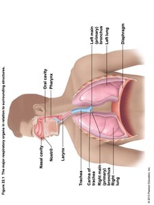
The respiratory system consists of several major organs, each with specialized roles in air conduction and gas exchange.
Major organs:
Nose, nasal cavity, and paranasal sinuses
Pharynx
Larynx
Trachea
Bronchi and their branches
Lungs and alveoli
Functional Anatomy: Zones of the Respiratory System
The respiratory system is divided into conducting and respiratory zones, each with distinct structures and functions.
Respiratory zone: Site of gas exchange; includes microscopic structures such as respiratory bronchioles, alveolar ducts, and alveoli.
Conducting zone: Conduits to gas exchange sites; includes all other respiratory structures, cleanses, warms, and humidifies air.
Diaphragm and other respiratory muscles: Promote ventilation by changing thoracic volume.
Upper Respiratory Tract: Nasal Cavity and Paranasal Sinuses
The upper respiratory tract conditions incoming air and provides resonance for speech.
Paranasal sinuses: Located in frontal, sphenoid, ethmoid, and maxillary bones.
Functions: Lighten the skull, secrete mucus, help warm and moisten air.
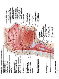
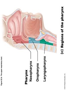
Pharynx
The pharynx is a muscular tube that serves as a passageway for both air and food, divided into three regions.
Regions:
Nasopharynx
Oropharynx
Laryngopharynx
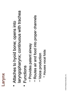
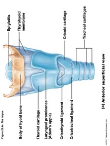
Larynx
The larynx, or voice box, is responsible for maintaining an open airway, routing food and air, and producing sound.
Attaches to the hyoid bone and opens into the laryngopharynx; continuous with the trachea.
Functions:
Provides a patent airway
Routes air and food into proper channels
Voice production (houses vocal folds)
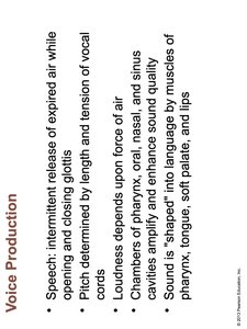
Voice Production
Voice is produced by the intermittent release of expired air through the glottis, causing the vocal folds to vibrate.
Pitch is determined by the length and tension of the vocal cords.
Loudness depends on the force of air across the cords.
Sound quality is shaped by the pharynx, oral, nasal, and sinus cavities, and articulated by the tongue, soft palate, and lips.
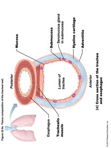
Trachea
The trachea, or windpipe, is a flexible tube supported by C-shaped cartilaginous rings, conducting air to the bronchi.
Composed of mucosa, submucosa, hyaline cartilage, and adventitia.
Lined with pseudostratified ciliated columnar epithelium and goblet cells for mucus production and debris removal.
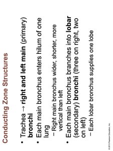
Conducting Zone Structures
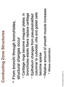
The conducting zone includes the trachea, bronchi, and bronchioles, which transport air to the respiratory zone.
Trachea divides into right and left main (primary) bronchi.
Main bronchi branch into lobar (secondary) bronchi, then into segmental (tertiary) bronchi, and finally into bronchioles.
Structural changes occur along the pathway: cartilage rings become plates, epithelium thins, and smooth muscle increases.
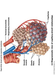
Respiratory Zone
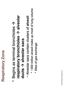
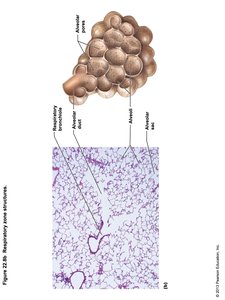
The respiratory zone is where gas exchange occurs, beginning at the respiratory bronchioles and ending at the alveolar sacs.
Respiratory bronchioles lead to alveolar ducts, which terminate in alveolar sacs composed of clusters of alveoli.
Alveoli are the primary sites of gas exchange; about 300 million alveoli make up most of the lung volume.
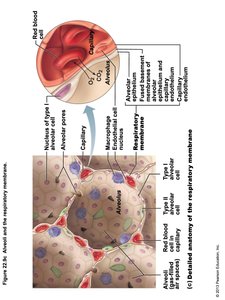
Respiratory Membrane
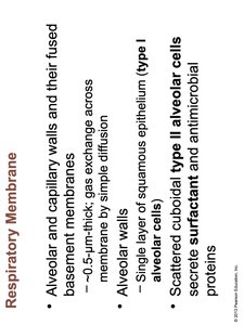
The respiratory membrane is the thin barrier through which gases are exchanged between alveolar air and blood in the capillaries.
Composed of alveolar and capillary walls and their fused basement membranes (~0.5 μm thick).
Alveolar walls consist of a single layer of squamous epithelium (type I alveolar cells).
Type II alveolar cells secrete surfactant and antimicrobial proteins.
Lungs and Pleurae
The lungs are paired organs in the thoracic cavity, each divided into lobes and surrounded by pleural membranes.
Right lung: three lobes; left lung: two lobes (to accommodate the heart).
Pleurae are serous membranes that reduce friction and compartmentalize the lungs.
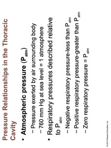
Pressure Relationships in the Thoracic Cavity
Ventilation depends on pressure differences between the atmosphere and the lungs, governed by Boyle's Law.
Atmospheric pressure (Patm): Pressure exerted by air surrounding the body (760 mm Hg at sea level).
Respiratory pressures are described relative to Patm:
Negative: less than Patm
Positive: greater than Patm
Zero: equal to Patm
Boyle's Law
Boyle's Law describes the inverse relationship between the pressure and volume of a gas at constant temperature.
As volume increases, pressure decreases, and vice versa.
Mathematically:
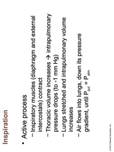
Mechanics of Breathing: Inspiration and Expiration
Breathing involves the movement of air into (inspiration) and out of (expiration) the lungs, driven by changes in thoracic volume and pressure.
Inspiration: Active process; diaphragm and external intercostal muscles contract, thoracic volume increases, intrapulmonary pressure drops, air flows in.
Forced inspiration: Accessory muscles (scalenes, sternocleidomastoid, pectoralis minor) further increase thoracic volume during vigorous exercise or respiratory distress.
Expiration: Normally passive; inspiratory muscles relax, thoracic volume decreases, intrapulmonary pressure rises, air flows out.
Forced expiration: Active process using abdominal and internal intercostal muscles.
Respiratory Volumes and Capacities
Respiratory volumes and capacities are measured to assess lung function and include tidal volume, inspiratory and expiratory reserve volumes, and vital capacity.
Tidal volume (TV): Amount of air inhaled or exhaled with each breath under resting conditions.
Inspiratory reserve volume (IRV): Amount of air that can be forcefully inhaled after a normal tidal volume inhalation.
Expiratory reserve volume (ERV): Amount of air that can be forcefully exhaled after a normal tidal volume exhalation.
Vital capacity (VC): Total amount of exchangeable air (TV + IRV + ERV).
Respiratory: Physiological Overview
Gas exchange and transport are critical physiological processes of the respiratory system, involving oxygen and carbon dioxide movement and regulation of breathing.
Gas exchange: Occurs by diffusion, dictated by concentration (partial pressure) gradients.
Transport of O2: Mostly bound to hemoglobin in red blood cells.
Transport of CO2: Primarily as bicarbonate ion (HCO3-), with some bound to hemoglobin or dissolved in plasma.
Control of respiration: Depth and rate are regulated by neural and chemical factors, including blood levels of CO2 and O2.
Additional info: The respiratory system is closely integrated with the cardiovascular system to ensure efficient gas transport and homeostasis. Disorders of the respiratory system can significantly impact oxygen delivery and acid-base balance.
