 Back
BackThe Respiratory System: Structure, Function, and Clinical Relevance
Study Guide - Smart Notes
 Tailored notes based on your materials, expanded with key definitions, examples, and context.
Chapter 22: The Respiratory System
Overview and Functions of the Respiratory System
The respiratory system is essential for gas exchange, speech, olfaction, and maintaining homeostasis. It consists of organs and structures that facilitate the movement of air and the exchange of oxygen (O2) and carbon dioxide (CO2) between the atmosphere and the bloodstream.
Gas Exchange: O2 is absorbed and CO2 is expelled between blood and air in the lungs.
Speech and Vocalization: Air movement through the larynx produces sound.
Olfaction: The nasal cavity houses olfactory receptors for the sense of smell.
pH Regulation: Removal of CO2 helps control blood pH.
Blood Pressure Regulation: The lungs produce angiotensin II, a vasoconstrictor.
Assists Circulation: Breathing aids venous and lymphatic return to the heart.
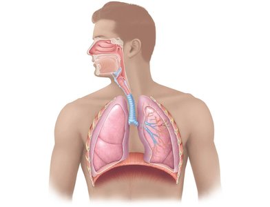
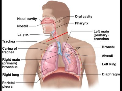
Anatomy of the Respiratory System
Major Divisions
Upper Respiratory Tract: Nose, nasal cavity, paranasal sinuses, pharynx, and larynx.
Lower Respiratory Tract: Trachea, bronchi, bronchioles, and lungs.
The Nose and Nasal Cavity
The nose provides an airway, moistens and warms air, filters particles, resonates sound, and houses olfactory receptors. The nasal cavity contains conchae (turbinate bones) that increase surface area and enhance air turbulence.
Olfactory mucosa: Contains smell receptors.
Respiratory mucosa: Filters, heats, and moistens air.
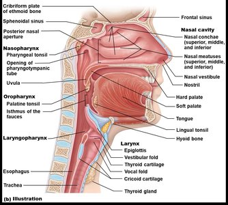
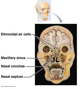
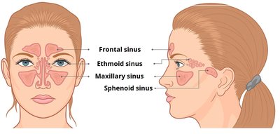
Paranasal Sinuses
Paranasal sinuses are air-filled spaces that open into the nasal cavity and are lined by respiratory mucosa. They lighten the skull, warm and moisten air, and contribute to voice resonance.
The Pharynx
The pharynx is a muscular tube that serves as a passageway for air and food. It is divided into three regions:
Nasopharynx: Lined with pseudostratified ciliated columnar epithelium; contains the pharyngeal tonsil and pharyngotympanic tube.
Oropharynx: Lined with nonkeratinized stratified squamous epithelium; contains palatine and lingual tonsils.
Laryngopharynx: Also lined with nonkeratinized stratified squamous epithelium; continuous with the esophagus and larynx.
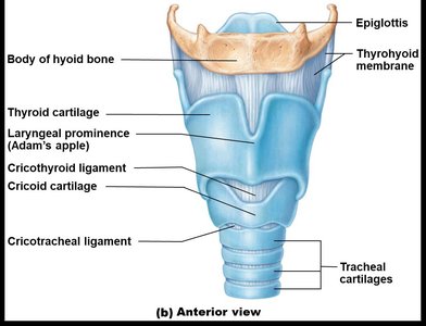
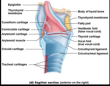
The Larynx
The larynx connects the pharynx to the trachea and is responsible for voice production, maintaining an open airway, and routing air and food using the epiglottis.
Cartilages: Includes thyroid, cricoid, arytenoid, and epiglottis.
Vocal Folds: True vocal cords produce sound; vestibular folds (false vocal cords) do not.
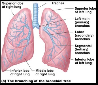
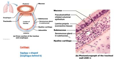
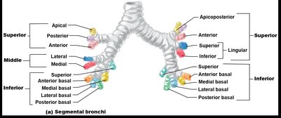
The Trachea and Bronchial Tree
The trachea is a flexible tube supported by C-shaped hyaline cartilage rings. It divides into the right and left main (primary) bronchi, which branch into secondary (lobar) and tertiary (segmental) bronchi, and then into bronchioles and terminal bronchioles.
Tracheal Wall: Lined with pseudostratified ciliated columnar epithelium and contains goblet cells.
Bronchial Tree: Extensive branching network that conducts air to the alveoli.
Histology of the Respiratory Tract
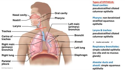
The respiratory tract is lined by different types of epithelium depending on the region:
Nasal cavity, larynx, trachea: Pseudostratified ciliated columnar epithelium.
Pharynx: Non-keratinized stratified squamous epithelium.
Bronchioles: Simple cuboidal epithelium.
Alveoli: Simple squamous epithelium (Type I alveolar cells).
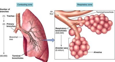
Conducting Zone vs. Respiratory Zone
The respiratory system is divided into two functional zones:
Conducting Zone: Includes all structures that transport air but do not participate in gas exchange (nasal cavity to terminal bronchioles).
Respiratory Zone: Includes respiratory bronchioles, alveolar ducts, and alveoli where gas exchange occurs.
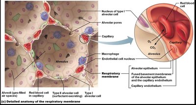
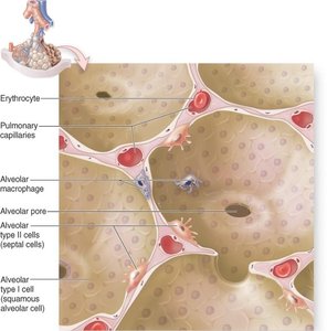
Alveoli and Gas Exchange
Alveoli are tiny air sacs where gas exchange occurs. They are surrounded by capillaries and have thin walls to facilitate diffusion of gases.
Type I alveolar cells: Simple squamous cells forming the respiratory membrane.
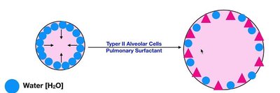
Type II alveolar cells: Secrete surfactant to reduce surface tension and prevent alveolar collapse.
Alveolar macrophages: Remove debris and pathogens.
Alveolar pores: Equalize air pressure and provide alternate air routes.
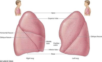
Major Landmarks of the Lungs
The lungs are divided into lobes and segments, each supplied by its own bronchus and blood vessels. The right lung has three lobes; the left lung has two lobes and a cardiac notch.
The Pleurae
The pleurae are serous membranes that surround each lung and line the thoracic cavity. They reduce friction and help maintain lung inflation.
Parietal pleura: Lines the thoracic wall.
Visceral pleura: Covers the lungs.
Pleural cavity: Contains lubricating fluid.
Respiratory Physiology and Terminology
Key Terms
Respiration: The overall process of gas exchange.
External Respiration: Gas exchange between air in the lungs and blood.
Internal Respiration: Gas exchange between blood and tissues.
Pulmonary Ventilation: Movement of air into (inspiration) and out of (expiration) the lungs.
Cellular Respiration: Metabolic processes (glycolysis, Krebs cycle, oxidative phosphorylation) that produce ATP.
CO2 Transport and Acid-Base Balance
CO2 is transported in the blood as dissolved gas, bound to hemoglobin, or as bicarbonate ions. The following equation summarizes the conversion of CO2 to bicarbonate, which is catalyzed by the enzyme carbonic anhydrase:
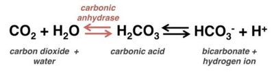
This reaction is crucial for maintaining blood pH and is reversed in the lungs to allow exhalation of CO2.
Summary Table: Conducting vs. Respiratory Zone Structures
Structure | Inner Diameter (mm) | Cilia | Goblet Cells | Cartilage | Smooth Muscle |
|---|---|---|---|---|---|
Larynx | 35–45 | +++ | +++ | +++ (plates) | + |
Trachea | 20–25 | +++ | +++ | +++ (C-shaped) | + |
Primary Bronchi | 12–16 | +++ | +++ | +++ (rings) | ++ |
Secondary Bronchi | 8–10 | +++ | +++ | ++ (plates) | ++ |
Tertiary Bronchi | 1–8 | +++ | +++ | + (plates) | +++ |
Smaller Bronchi | 1–8 | +++ | +++ | + (plates) | +++ |
Terminal Bronchioles | <0.5 | + | 0 | 0 | +++ |
Respiratory Bronchioles | <0.5 | 0 | 0 | 0 | ++ |
Alveolar Sacs | 0 | 0 | 0 | 0 | 0 |
Additional info: Table summarizes the structural and functional differences between the conducting and respiratory zones.
Key Concepts for Exam Preparation
Know the anatomical divisions and histology of the respiratory tract.
Understand the functional differences between the conducting and respiratory zones.
Be able to describe the process of gas exchange and the role of alveolar cells.
Explain the importance of surfactant and the acid-base balance equation in respiratory physiology.
