 Back
BackThe Respiratory System: Structure, Function, and Clinical Conditions
Study Guide - Smart Notes
 Tailored notes based on your materials, expanded with key definitions, examples, and context.
The Respiratory System
Overview and Structural Plan
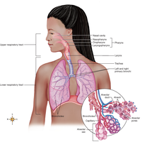
The respiratory system is essential for gas exchange, supplying oxygen to the body and removing carbon dioxide. Its structure resembles an inverted tree, with the trachea as the trunk and the alveoli as the leaves, where gas exchange occurs via diffusion.
Major Functions: Air distribution, gas exchange, air purification, and voice production.
Diffusion: The primary mechanism for gas exchange between alveoli and capillaries.
Respiratory Tracts
The respiratory system is divided into upper and lower tracts, each with distinct structures and functions.
Upper Respiratory Tract: Nose, pharynx, larynx
Lower Respiratory Tract: Trachea, bronchial tree, lungs
Clinical Comparison: A "head cold" affects the upper tract (nasal congestion, sore throat), while a "chest cold" affects the lower tract (dyspnea, cough).
Respiratory Mucosa
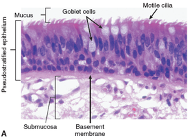
The respiratory mucosa is a mucous membrane lining the airways, producing over 125 mL of mucus daily to trap irritants. Cilia move mucus upward to the pharynx for removal, serving as an air purification mechanism.
Respiratory Membrane: Barrier between alveoli and capillaries for gas exchange.
Respiratory Mucosa: Lines the respiratory tract and secretes mucus for protection.
Nose and Paranasal Sinuses
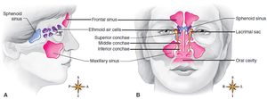
The nose is divided by the nasal septum and lined with mucous membrane. Paranasal sinuses (frontal, maxillary, sphenoidal, ethmoidal) drain into the nose and contribute to voice resonance.
Olfactory Receptors: Located in the nasal mucosa, responsible for the sense of smell.
Sinus Function: Affect voice resonance and humidify inhaled air.
Lacrimal Pathway: Tears drain from lacrimal sacs into the nasal cavity.
Pharynx (Throat)
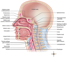
The pharynx is a 12.5 cm tube divided into nasopharynx, oropharynx, and laryngopharynx. It serves as a passageway for both air (respiratory system) and food/liquids (digestive system).
Tonsils: Lymphoid tissue in the pharynx providing immune protection.
Eustachian Tube: Equalizes pressure in the middle ear.
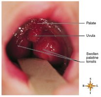
Clinical Note: Swollen adenoids can cause dyspnea; tonsillectomy removes chronically infected tissue but also immune tissue.
Tonsillitis
Tonsillitis is the inflammation of the tonsils, often resulting in sore throat and difficulty swallowing. Tonsils are composed of lymphoid tissue and play a role in immune defense.
Larynx (Voice Box)
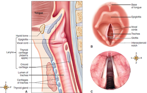
The larynx is located below the pharynx and is composed of several cartilages, including the thyroid cartilage (Adam's apple). The epiglottis covers the laryngeal opening during swallowing to prevent aspiration. Vocal cords within the larynx produce sound as air passes over them.
Function: Air passage and voice production.
Clinical Note: Laryngeal cancer is more common in older men and those with alcohol abuse; removal of the larynx requires alternative speech methods.
Trachea (Windpipe)
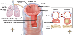
The trachea is an 11 cm tube supported by C-shaped cartilage rings, ensuring it remains open for air passage. Its mucous lining and ciliated epithelium help remove contaminants.
Obstruction: Blockage can be fatal; abdominal thrust maneuver or tracheostomy may be required.
Bronchi, Bronchioles, and Alveoli
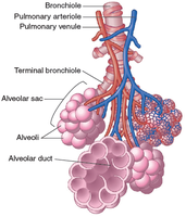
The trachea divides into right and left primary bronchi, which branch into smaller bronchi and bronchioles, ending in alveolar sacs. The right bronchus is more vertical, making it more likely for aspirated objects to lodge there. Alveoli are the primary sites for gas exchange, and type II cells produce surfactant to reduce surface tension.
Function: Air distribution and gas exchange.
Surfactant: Prevents alveolar collapse by reducing surface tension.
Summary Table: Major Respiratory Structures and Functions
Structure | Main Function |
|---|---|
Nose | Warms, moistens, and filters air; sense of smell |
Pharynx | Passageway for air and food; immune defense (tonsils) |
Larynx | Voice production; air passage |
Trachea | Air passage; removal of contaminants |
Bronchi/Bronchioles | Air distribution |
Alveoli | Gas exchange |
Clinical Conditions of the Respiratory System
Upper Respiratory Infections (URIs): Rhinitis, pharyngitis, laryngitis, epiglottitis, croup
Anatomical Conditions: Deviated septum, epistaxis (nosebleed)
Lower Respiratory Conditions: Acute bronchitis, pneumonia, tuberculosis, COPD, asthma, lung cancer
Respiratory Distress Syndromes: IRDS (infants, lack of surfactant), ARDS (adults, surfactant impairment)
Mechanics of Breathing (Pulmonary Ventilation)
Breathing consists of inspiration (active, diaphragm and external intercostals contract) and expiration (passive, elastic recoil; active during forceful expiration with internal intercostals and abdominal muscles). Air moves due to pressure gradients created by changes in thoracic volume.
Key Equation: As volume increases, pressure decreases, and vice versa.
Pulmonary Volumes
Tidal Volume (TV): Normal breath (~500 mL)
Vital Capacity (VC): Maximum air exhaled after maximum inhalation
Expiratory Reserve Volume (ERV): Extra air exhaled after normal expiration
Inspiratory Reserve Volume (IRV): Extra air inhaled after normal inspiration
Residual Volume (RV): Air remaining after forceful expiration
Regulation of Ventilation
Breathing is regulated by respiratory centers in the brainstem (medulla and pons). Chemoreceptors monitor CO2, O2, and pH levels, while stretch receptors prevent overinflation. The cerebral cortex allows voluntary control, but this is limited.
Medullary Centers: Set basic rhythm (12–18 breaths/min)
Pontine Centers: Modify rhythm as needed
Breathing Patterns
Eupnea: Normal breathing
Hyperventilation: Rapid, deep breathing
Hypoventilation: Slow, shallow breathing
Dyspnea: Difficult breathing
Orthopnea: Dyspnea relieved by sitting upright
Apnea: Absence of breathing
Cheyne-Stokes Respiration: Alternating apnea and hyperventilation
Gas Exchange and Transport
Gas exchange occurs in the alveoli (external respiration) and at the tissues (internal respiration). Oxygen is transported mainly as oxyhemoglobin, while carbon dioxide is transported as dissolved CO2, carbaminohemoglobin, and bicarbonate ions.
Oxygen Transport: Mostly as oxyhemoglobin (HbO2)
Carbon Dioxide Transport: 10% dissolved, 20% as carbaminohemoglobin, 70% as bicarbonate (HCO3−)
Additional info: The respiratory system is closely integrated with the cardiovascular system for efficient gas transport and exchange. Disorders affecting one system often impact the other.
