 Back
BackThe Respiratory System: Part 2 AP 2nd year Lungs
Study Guide - Smart Notes
 Tailored notes based on your materials, expanded with key definitions, examples, and context.
The Trachea
Structure and Function
The trachea, commonly known as the windpipe, is a flexible tube that extends from the larynx into the mediastinum, where it divides into the two main bronchi. It is approximately 4 inches in length and 3/4 inch in diameter. The trachea serves as the main passageway for air to enter the lungs.
Location: Extends from the larynx to the mediastinum, dividing into right and left main bronchi.
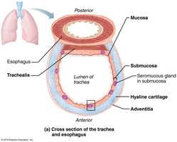
Structure: Flexible, supported by C-shaped hyaline cartilage rings that keep the airway open.
Function: Conducts air to the bronchi; its ciliated mucosa helps filter and expel debris.
Histological Layers of the Trachea
The tracheal wall is composed of several distinct layers, each with specialized functions:
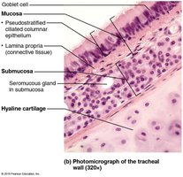
Mucosa: Ciliated pseudostratified epithelium with goblet cells that secrete mucus to trap particles.
Submucosa: Connective tissue layer containing seromucous glands and supported by hyaline cartilage rings.
Adventitia: Outermost connective tissue layer that anchors the trachea.
Specialized Structures
Trachealis Muscle: Smooth muscle that connects the posterior parts of the cartilage rings; contracts during coughing to expel mucus.
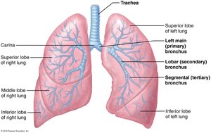
Carina: The last tracheal cartilage at the point where the trachea branches into the main bronchi; highly sensitive and triggers violent coughing if contacted by foreign objects.
Bronchi and the Bronchial Tree
Branching Pattern
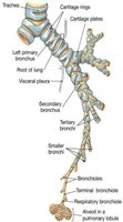
The bronchial tree refers to the extensive branching of air passages within the lungs, undergoing approximately 23 orders of branching from the trachea to the alveoli.
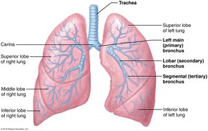
Main (Primary) Bronchi: The trachea divides into right and left main bronchi. The right bronchus is wider, shorter, and more vertical than the left.
Lobar (Secondary) Bronchi: Each main bronchus branches into lobar bronchi—three on the right, two on the left—each supplying a lung lobe.
Segmental (Tertiary) Bronchi: Each lobar bronchus divides into segmental bronchi, which continue to branch into smaller bronchioles.
Bronchioles: Airways less than 1 mm in diameter; terminal bronchioles are less than 0.5 mm in diameter.
Structural Changes Along the Conducting Zone
Support Structures: Cartilage rings become irregular plates and are eventually replaced by elastic fibers in bronchioles.
Epithelium: Changes from pseudostratified columnar to cuboidal; cilia and goblet cells become sparse.
Smooth Muscle: Increases as airways become smaller, allowing bronchioles to regulate airflow resistance.
Respiratory Zone Structures
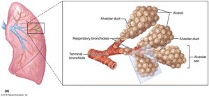
The respiratory zone begins at the junction of terminal bronchioles and respiratory bronchioles, leading to alveolar ducts and alveolar sacs. Alveolar sacs contain clusters of alveoli, which are the primary sites of gas exchange.
Alveoli: Approximately 300 million in the lungs, providing a large surface area for gas exchange.
Respiratory Membrane and Alveoli
Structure and Function
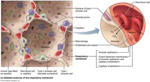
The respiratory membrane is the thin barrier through which gas exchange occurs between alveolar air and blood in pulmonary capillaries.
Components: Alveolar wall (simple squamous epithelium, type I cells), capillary wall, and fused basement membranes.
Type II Alveolar Cells: Scattered cuboidal cells that secrete surfactant to reduce surface tension.
Alveolar Macrophages: Immune cells that keep alveolar surfaces sterile.
Alveolar Pores: Connect adjacent alveoli, providing alternate routes for air in case of blockages.
Lung Anatomy
Gross Structure
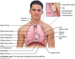
The lungs occupy the thoracic cavity except for the mediastinum. Each lung has an apex (superior tip) and a base (rests on the diaphragm).
Hilum: The mediastinal surface where blood vessels, bronchi, lymphatic vessels, and nerves enter and exit the lung.
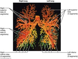
Lobes: The right lung has three lobes (superior, middle, inferior), while the left lung has two (superior, inferior) and a cardiac notch.
Bronchopulmonary Segments and Lobules
Bronchopulmonary Segments: Each lobe is divided into segments (10 on right, 8-10 on left), each served by its own artery, vein, and tertiary bronchus.
Lobules: Smallest subdivisions visible to the naked eye, hexagonal in shape, served by bronchioles and branches.
Stroma: The elastic connective tissue that makes the lungs spongy and elastic.
Blood Supply of the Lungs
Pulmonary and Bronchial Circulation
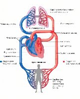
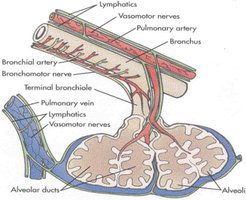
Pulmonary Circulation: Pulmonary arteries deliver systemic venous blood to the lungs for oxygenation; pulmonary veins return oxygenated blood to the heart. This is a low-pressure, high-volume system.
Bronchial Circulation: Bronchial arteries (from the aorta) provide oxygenated blood to lung tissue. This is a high-pressure, low-volume system. Bronchial veins merge with pulmonary veins to return blood to the heart.
Innervation of the Lungs
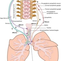
The lungs are innervated by both the parasympathetic and sympathetic nervous systems, as well as visceral sensory fibers.
Parasympathetic Stimulation: Causes bronchoconstriction (narrowing of airways).
Sympathetic Stimulation: Causes bronchodilation (widening of airways).
Pleurae
Structure and Function
The pleurae are thin, double-layered serosal membranes surrounding each lung and lining the thoracic cavity.
Parietal Pleura: Lines the thoracic wall, diaphragm, and mediastinum.
Visceral Pleura: Covers the external lung surface.
Pleural Fluid: Fills the pleural cavity, providing lubrication and surface tension for lung expansion and recoil.
Respiratory Physiology
Breathing Mechanics
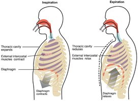
Pulmonary ventilation consists of two phases: inspiration (air flows into the lungs) and expiration (air exits the lungs). These processes depend on changes in thoracic cavity volume and pressure relationships.
Inspiration: Diaphragm contracts, thoracic cavity expands, and air flows in.
Expiration: Diaphragm relaxes, thoracic cavity volume decreases, and air is expelled.
Pressure Relationships in the Thoracic Cavity
Atmospheric Pressure (Patm): The pressure exerted by air surrounding the body, typically 760 mmHg at sea level (1 atmosphere).
Respiratory Pressures: Expressed relative to Patm. Negative pressure is less than Patm, positive is greater, and zero is equal to Patm.
