 Back
The Senses – Vision: Anatomy and Physiology of the Eye
Study Guide - Smart Notes
 Tailored notes based on your materials, expanded with key definitions, examples, and context.
Introduction to Vision
Vision is one of the five special senses and is essential for interacting with our environment. The eye, along with its accessory structures, enables the detection and processing of light, allowing us to perceive the world around us. This section covers the anatomy of the eye, its accessory structures, and the physiological processes involved in vision.
Sensation vs. Perception
Definitions and Processes
Sensation: The activation of sensory receptor cells by a stimulus, leading to the generation of action potentials in sensory neurons.
Perception: The conscious interpretation of sensory information, which occurs in the sensory cortices of the cerebrum.
Sensation is necessary for perception, but not all sensations reach conscious awareness.
These principles apply to all sensory modalities, including vision, hearing, taste, smell, and touch.
Accessory Structures of the Eye
Protection and Support
The orbit is a bony cavity in the skull that houses and protects the eyeball.
Eyelids and eyelashes protect the eye from debris and injury.
The conjunctiva is a thin membrane lining the inner eyelids (palpebral conjunctiva) and covering the white of the eye (ocular conjunctiva).
The lacrimal gland produces tears that clean and nourish the cornea, draining through the lacrimal ducts and nasolacrimal duct into the nasal cavity.
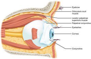
Muscles of the Eye
Extraocular Muscles and Eye Movement
Six extraocular muscles control eye movement: four rectus muscles (superior, inferior, medial, lateral) and two oblique muscles (superior, inferior).
The rectus muscles move the eye up, down, medially, and laterally.
The oblique muscles rotate the eye and compensate for the angle of the rectus muscles.
Innervation: Lateral rectus (abducens nerve), superior oblique (trochlear nerve), others (oculomotor nerve).
Anatomy of the Eyeball
Layers of the Eyeball
Fibrous tunic: Outermost layer; includes the white sclera and transparent cornea.
Vascular tunic: Middle layer; includes the choroid (vascular supply), ciliary body (lens accommodation), and iris (controls pupil size).
Neural tunic (retina): Innermost layer; contains photoreceptors and neural tissue for photoreception.
Cavities of the Eyeball
Anterior cavity: Between cornea and lens; contains aqueous humor and is divided into anterior and posterior chambers.
Posterior cavity: Behind the lens; filled with vitreous humor, a gel-like substance that maintains eye shape.
The Retina
Structure and Function
The retina contains two types of photoreceptors: rods (sensitive to low light, peripheral vision) and cones (color vision, high acuity).
Rods contain the pigment rhodopsin; cones contain opsins sensitive to red, green, or blue light.
Photoreceptors synapse with bipolar cells, which in turn synapse with ganglion cells. The axons of ganglion cells form the optic nerve.
The fovea centralis is the region of highest visual acuity, where light directly reaches cones.
The optic disc is a blind spot where the optic nerve exits the eye; no photoreceptors are present here.
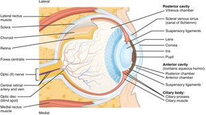
Pathway of Light Through the Eye
Refraction and Focusing
Light enters through the cornea, passes through the aqueous humor, pupil, lens, and vitreous humor before reaching the retina.
The cornea and lens refract (bend) light to focus it on the retina.
The iris adjusts pupil size to regulate the amount of light entering the eye.
Dissection of the Cow Eye
Observation and Structure-Function Relationships
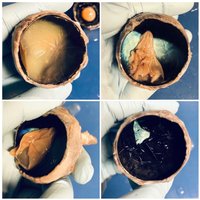
The cow eye is structurally similar to the human eye and is used for anatomical study.
Key structures observed include the sclera, cornea, muscles, fat, pupil, iris, lens, optic nerve, aqueous humor, vitreous humor, retina, and tapetum lucidum (reflective layer not present in humans).
The tapetum lucidum enhances night vision in animals by reflecting light back through the retina.
Summary Table: Major Eye Structures and Functions
Structure | Function |
|---|---|
Sclera | Protects and maintains the shape of the eyeball |
Cornea | Refracts light entering the eye |
Iris | Controls the diameter of the pupil |
Pupil | Regulates the amount of light entering the eye |
Lens | Focuses light onto the retina |
Retina | Contains photoreceptors for vision |
Optic nerve | Transmits visual information to the brain |
Vitreous humor | Maintains eye shape and optical properties |
Aqueous humor | Nourishes cornea and lens; maintains intraocular pressure |
Tapetum lucidum (cow only) | Reflects light to enhance night vision |
Sample Questions and Answers
What is the major anatomical difference between the cow eye and human eye? The cow eye contains a tapetum lucidum, which is absent in humans.
Why does the optic nerve cause a blind spot? There are no photoreceptors at the optic disc where the optic nerve exits the eye.
What happens to the pupil in a dark room? The pupil dilates (gets larger) to allow more light to enter the eye.
What is the function of the muscles surrounding the eye? They move the eyeball, allowing us to direct our gaze and focus on objects of interest.
Does light first enter the eye through the lens? False. Light first enters through the cornea.
What is the white layer that surrounds the eye? The sclera.
Order of parts through which light passes: Cornea, lens, vitreous humor, posterior cavity.
Key Equations and Concepts
Refraction of Light: The bending of light as it passes through different media (cornea, aqueous humor, lens, vitreous humor).
Accommodation: The process by which the ciliary body changes the shape of the lens to focus light on the retina.
Equation for lens focusing (thin lens equation):
Where is the focal length, is the object distance, and is the image distance.
Additional info: The tapetum lucidum is a reflective layer found in the eyes of many vertebrates (not humans) that enhances night vision by reflecting light back through the retina, increasing the light available to photoreceptors.
