 Back
BackThe Skeletal System: Bone Tissue – Structure, Function, and Physiology
Study Guide - Smart Notes
 Tailored notes based on your materials, expanded with key definitions, examples, and context.
The Skeletal System: Bone Tissue
Introduction to Bone Tissue
Bone is a dynamic organ composed of several tissue types, including bone tissue, cartilage, dense connective tissue, epithelium, blood-forming tissues, adipose tissue, and nervous tissue. Each bone is considered an organ and is constantly remodeled throughout life. Together, bones and their associated cartilages form the skeletal system.
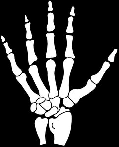
Support and Protection: Bones provide structural support and protect soft tissues.
Movement: Serve as attachment sites for muscles, enabling movement.
Mineral Storage: Store calcium and phosphate, maintaining mineral homeostasis.
Blood Cell Production: Red bone marrow produces blood cells (hemopoiesis).
Energy Storage: Yellow bone marrow stores fat as an energy reserve.
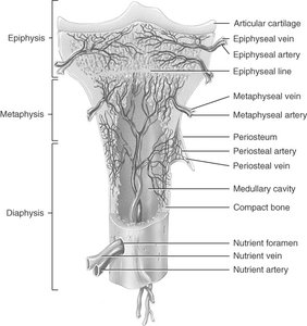
Anatomy of a Long Bone
Structural Regions
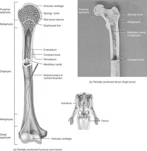
Long bones have distinct anatomical regions that contribute to their function and growth.
Diaphysis: The shaft or central part of the bone.
Epiphysis: The ends of the bone, typically expanded for joint articulation.
Metaphysis: The region between the diaphysis and epiphysis, containing the epiphyseal plate in growing bones.
Articular Cartilage: Covers joint surfaces, reducing friction and absorbing shock.
Medullary Cavity: Central cavity containing bone marrow.
Endosteum: Membrane lining the medullary cavity.
Periosteum: Tough outer membrane (fibrous and osteogenic layers) covering bone except at joint surfaces.
Histology of Bone Tissue
Bone Matrix and Cellular Components
Bone tissue is a specialized connective tissue with widely spaced cells embedded in a matrix. The matrix consists of 25% water, 25% collagen fibers, and 50% crystallized mineral salts, primarily hydroxyapatite and calcium carbonate. The process of calcification deposits these minerals in a collagen framework, giving bone its hardness and tensile strength.
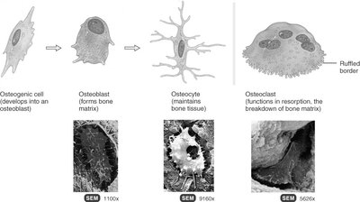
Osteoprogenitor (Osteogenic) Cells: Stem cells that differentiate into osteoblasts; found in periosteum and endosteum.
Osteoblasts: Bone-forming cells that secrete matrix but do not divide.
Osteocytes: Mature bone cells maintaining bone tissue.
Osteoclasts: Large cells derived from monocytes, responsible for bone resorption.
Types of Bone Tissue
Compact Bone
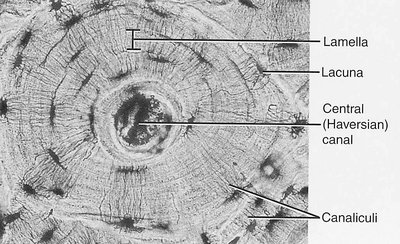
Compact bone is organized into osteons (Haversian systems), which contain concentric lamellae of calcified matrix surrounding a central canal with blood vessels and nerves. Osteocytes reside in lacunae and communicate via canaliculi. Compact bone forms the external layer of all bones and the shaft of long bones, providing strength to resist stress.
Spongy Bone
Spongy (cancellous) bone lacks osteons and consists of trabeculae, which are oriented along lines of stress. The spaces between trabeculae are filled with red bone marrow, supporting hematopoiesis. Spongy bone is lighter and found in short, flat, irregular bones, and the epiphyses of long bones.
Blood Supply of Bone
Bones are richly vascularized. Blood supply comes from periosteal arteries (supplying the periosteum), nutrient arteries (entering through nutrient foramina to supply the diaphysis and marrow), and metaphyseal and epiphyseal arteries (supplying the ends of bones).
Bone Formation (Ossification)
Overview
Bone formation, or ossification, occurs by two mechanisms: intramembranous and endochondral ossification. All embryonic connective tissue begins as mesenchyme.
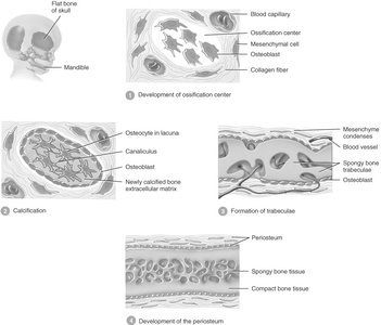
Intramembranous Ossification: Bone develops directly from mesenchymal tissue; forms flat bones of the skull and mandible.
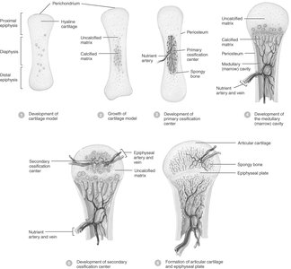
Endochondral Ossification: Bone develops by replacing hyaline cartilage; forms most bones of the body.
Intramembranous Ossification
Mesenchymal cells differentiate into osteoblasts, forming an ossification center.
Osteoblasts secrete osteoid, which calcifies and traps the cells, converting them into osteocytes.
Trabeculae form and fuse, creating spongy bone with red marrow.
Peripheral mesenchyme forms the periosteum.
Endochondral Ossification
Mesenchymal cells form a cartilage model, which grows in length (interstitial) and width (appositional).
Chondrocytes in the midregion die, triggering calcification.
Nutrient artery invades, osteoblasts and osteoclasts migrate, and primary ossification center forms in the diaphysis.
Secondary ossification centers develop in the epiphyses around birth; articular cartilage and epiphyseal plate remain.
Bone Growth
Growth in Length
Bones grow in length at the epiphyseal (growth) plate, which consists of four zones:
Zone of Resting Cartilage: Anchors plate to bone.
Zone of Proliferating Cartilage: Rapid cell division.
Zone of Hypertrophic Cartilage: Enlarged cells in columns.
Zone of Calcified Cartilage: Matrix calcifies, cells die, osteoclasts remove matrix, and osteoblasts form bone.
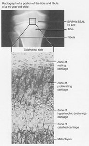
Growth in length ceases when the epiphyseal plate closes (epiphyseal line), typically between ages 18-25.

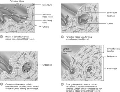
Growth in Thickness
Bones grow in thickness by appositional growth at the surface. Periosteal cells differentiate into osteoblasts, forming bony ridges and tunnels around blood vessels. Concentric lamellae fill in the tunnel, forming new osteons.
Factors Affecting Bone Growth
Nutrition: Adequate minerals (calcium, phosphorus) and vitamins (C, K, B12) are essential.
Hormones: Insulin-like growth factor, growth hormone, thyroid hormones, and sex hormones regulate growth and development.
Hormonal imbalances can lead to disorders such as gigantism, dwarfism, or abnormal growth plate closure.
Bone Remodeling
Bone remodeling is the continuous replacement of old bone by new bone tissue. Osteoclasts resorb bone, while osteoblasts form new bone. Remodeling is influenced by mechanical stress and hormones, allowing bones to adapt to new stresses and repair microdamage.
Fractures and Bone Repair
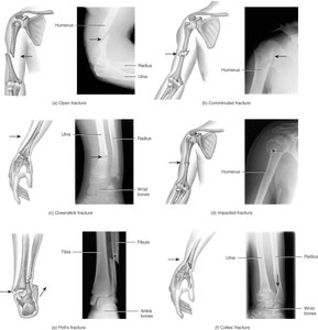
Types of Fractures
Greenstick: Partial fracture, common in children.
Impacted: One fragment driven into another.
Closed (Simple): No break in skin.
Open (Compound): Bone pierces the skin.
Comminuted: Bone fragments into several pieces.
Pott’s: Distal fibula/tibia fracture.
Colles’s: Distal radius/ulna fracture.
Stress Fracture: Microscopic fissures from repetitive stress.
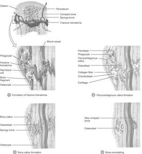
Bone Repair Process
Fracture Hematoma Formation: Blood clot forms, inflammation and phagocytosis occur.
Fibrocartilaginous (Soft) Callus Formation: Fibroblasts and chondroblasts produce collagen and cartilage to bridge the gap.
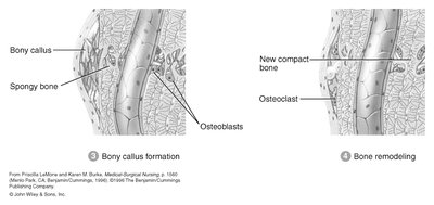
Bony (Hard) Callus Formation: Osteoblasts produce spongy bone, uniting the fragments.
Bone Remodeling: Compact bone replaces spongy bone, restoring normal shape.
Calcium Homeostasis and Bone Tissue
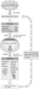
The skeleton acts as a reservoir for calcium and phosphate. Calcium is vital for nerve and muscle function, blood clotting, and enzyme activity. Blood calcium levels are tightly regulated (9–11 mg/100 mL); deviations can be life-threatening.
Parathyroid Hormone (PTH): Increases blood calcium by stimulating osteoclasts, enhancing kidney reabsorption, and promoting calcitriol production.
Calcitonin: Lowers blood calcium by inhibiting osteoclasts and stimulating osteoblasts.
Exercise, Aging, and Bone Disorders
Exercise and Bone Tissue
Mechanical stress increases bone strength by stimulating mineral deposition and collagen production. Lack of stress (e.g., immobilization, weightlessness) leads to bone loss.
Aging and Bone Tissue
Aging leads to decreased bone mass (osteoporosis) and reduced protein synthesis, making bones brittle and prone to fracture. These effects are more pronounced in postmenopausal women due to decreased estrogen.
Osteoporosis
Osteoporosis is characterized by decreased bone mass and increased fracture risk. Risk factors include age, gender, genetics, lifestyle, and hormonal status. Prevention includes adequate nutrition, weight-bearing exercise, and, for some, hormone replacement therapy.
Disorders of Bone Ossification
Rickets: In children, improper deposition of calcium salts leads to soft, deformed bones; caused by vitamin D deficiency.
Osteomalacia: In adults, new bone fails to ossify during remodeling, leading to bone pain and fractures; also due to vitamin D deficiency.
