 Back
The Skeletal System: Structure, Classification, and Bone Markings
Study Guide - Smart Notes
 Tailored notes based on your materials, expanded with key definitions, examples, and context.
The Skeletal System
Overview of the Skeletal System
The skeletal system is a complex framework of bones, joints, cartilages, and ligaments that provides structural support, protection, and movement for the human body. It is divided into two main subdivisions: the axial skeleton and the appendicular skeleton.
Axial skeleton: Includes the skull, vertebral column, and thoracic cage.
Appendicular skeleton: Comprises the limbs and girdles (pectoral and pelvic) that attach them to the axial skeleton.
Functions of Bones
Major Functions
Bones serve several essential functions in the body:
Support: Provide a rigid framework that supports the body and cradles soft organs.
Protection: Protect vital organs (e.g., skull protects the brain, rib cage protects thoracic organs).
Movement: Serve as levers for muscles to produce movement.
Mineral and Fat Storage: Store minerals such as calcium and phosphorus, and fats in the marrow cavities.
Blood Cell Formation (Hematopoiesis): Occurs in red bone marrow.
Classification of Bones
Types of Bone Tissue
Bones are composed of two main types of osseous tissue:
Compact bone: Dense, smooth, and homogeneous tissue forming the outer layer of bones.
Spongy bone: Consists of small needlelike pieces called trabeculae and many open spaces.
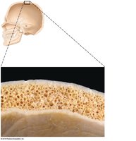
Bone Shapes
Bones are classified by shape into four groups:
Long bones: Longer than they are wide; mostly compact bone (e.g., femur, humerus).
Flat bones: Thin, flattened, usually curved; two layers of compact bone with spongy bone in between (e.g., skull, ribs, sternum).
Short bones: Cube-shaped; mostly spongy bone (e.g., carpals, tarsals).
Irregular bones: Complex shapes that do not fit other categories (e.g., vertebrae, hip bones).
Structure of Bone
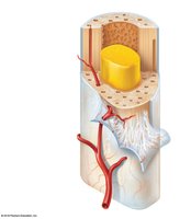
Gross Anatomy of a Long Bone
Long bones have a characteristic structure that supports their function:
Diaphysis: Shaft, composed of compact bone.
Epiphyses: Ends of the bone, mostly spongy bone covered by a thin layer of compact bone.
Periosteum: Fibrous connective tissue membrane covering the diaphysis, secured by Sharpey's fibers.
Articular cartilage: Hyaline cartilage covering epiphyses, reduces friction at joints.
Epiphyseal plate/line: Growth plate of hyaline cartilage in children; becomes the epiphyseal line in adults.
Medullary cavity: Central cavity in the diaphysis; contains yellow marrow (fat) in adults and red marrow (blood cell formation) in children.
Endosteum: Membrane lining the inner surface of the shaft.
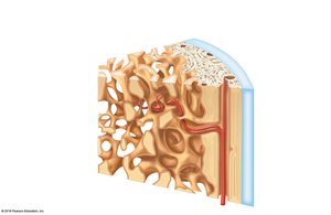
Microscopic Anatomy of Bone
Bone tissue is organized into structural units for strength and nutrient delivery:
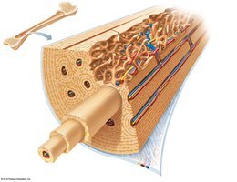
Osteon (Haversian system): The structural and functional unit of compact bone, consisting of concentric lamellae around a central canal.
Lamellae: Rings of bone matrix within an osteon.
Lacunae: Small cavities housing osteocytes (mature bone cells).
Canaliculi: Tiny canals connecting lacunae, allowing for nutrient and waste exchange.
Perforating (Volkmann's) canals: Canals perpendicular to the central canal, carrying blood vessels and nerves.
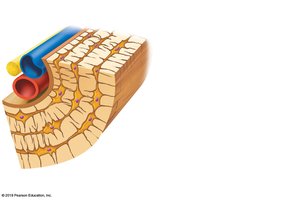
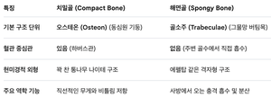
Comparison of Compact and Spongy Bone
특징 | 치밀골 (Compact Bone) | 해면골 (Spongy Bone) |
|---|---|---|
기본 구조 단위 | 오스테온 (Osteon) | 골소주 (Trabeculae) |
혈관 중심관 | 있음 (하버스관) | 없음 (주변 골수에서 직접 흡수) |
현미경적 형태 | 각 한 동나무 나이테 구조 | 여러방향 같은 격자형 구조 |
주요 역할 기능 | 직선적인 압력과 비틀림 저항 | 사방에서 오는 충격 흡수 및 분산 |
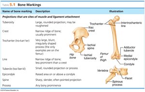
Bone Markings
Types and Functions of Bone Markings
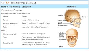
Bone markings are specific features on bones that serve as sites for muscle, tendon, and ligament attachment, or as passages for nerves and blood vessels. They are categorized as projections (processes) or depressions (cavities).
Name of bone marking | Description | Illustration |
|---|---|---|
Tuberosity | Large, rounded projection; may be roughened | Femur (intertrochanteric line) |
Crest | Narrow ridge of bone; usually prominent | Iliac crest |
Trochanter | Very large, blunt, irregularly shaped process (only on femur) | Femur (greater trochanter) |
Line | Narrow ridge of bone; less prominent than a crest | Femur (linea aspera) |
Tubercle | Small, rounded projection or process | Humerus (greater tubercle) |
Epicondyle | Raised area on or above a condyle | Femur (medial epicondyle) |
Spine | Sharp, slender, often pointed projection | Vertebra (spinous process) |
Process | Any bony prominence | Vertebra (transverse process) |
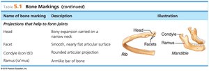
Name of bone marking | Description | Illustration |
|---|---|---|
Head | Bony expansion carried on a narrow neck | Femur head |
Facet | Smooth, nearly flat articular surface | Vertebra facet |
Condyle | Rounded articular projection | Mandible condyle |
Ramus | Arm-like bar of bone | Mandible ramus |
Name of bone marking | Description | Illustration |
|---|---|---|
Groove | Furrow | Skull groove |
Fissure | Narrow, slitlike opening | Skull fissure |
Foramen | Round or oval opening through a bone | Skull foramen |
Notch | Indentation at the edge of a structure | Mandible notch |
Meatus | Canal or tunnel-like passageway | Skull meatus |
Sinus | Cavity within a bone, filled with air and lined with mucous membrane | Skull sinus |
Fossa | Shallow, basinlike depression in a bone, often serving as an articular surface | Skull fossa |
Additional info:
Bones are organs composed of not only osseous tissue but also fibrous tissue, cartilage, adipose tissue, and blood.
Bone markings are essential for understanding muscle attachment, joint formation, and passage of nerves and vessels.
