 Back
BackThe Skeletal System: Structure, Function, and Physiology
Study Guide - Smart Notes
 Tailored notes based on your materials, expanded with key definitions, examples, and context.
The Skeletal System
Components of the Skeletal System
The skeletal system is composed of four main components: bone, cartilage, tendons, and ligaments. Each plays a distinct role in supporting and facilitating movement in the body.
Bone: Includes spongy and compact types; provides structural support and protection.
Cartilage: Types include hyaline, fibrocartilage, and elastic; cushions joints and supports soft tissues.
Tendons: Dense regular connective tissue; connects muscle to bone.
Ligaments: Dense regular connective tissue; connects bone to bone.
Functions of the Skeletal System
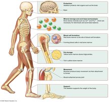
The skeletal system serves several essential functions for the human body, including support, protection, movement, mineral storage, blood cell production, and fat storage.
Support: Provides the structural framework and supports the body's weight.
Protection: Shields vital organs (e.g., cranium protects the brain, ribs protect the heart and lungs).
Movement: Muscles attach to bones; contraction pulls on bones to produce movement.
Mineral Homeostasis: Stores minerals such as calcium (99% of body’s content) and phosphorus.
Blood Cell Production: Red bone marrow produces red and white blood cells and platelets (hemopoiesis).
Fat Storage: Yellow bone marrow stores triglycerides as a chemical energy reserve.
Classification of Bones
Types of Bones
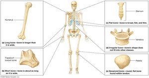
Bones are classified based on their shape and structure. The main types include long, short, flat, irregular, and sesamoid bones.
Long Bones: Longer than they are wide (e.g., humerus).
Short Bones: Nearly equal in length and width (e.g., trapezium).
Flat Bones: Thin, flat, and often curved (e.g., sternum).
Irregular Bones: Complex shapes that do not fit other categories (e.g., vertebra).
Sesamoid Bones: Small, round bones found within tendons (e.g., patella).
Structure of Long Bones
External and Internal Features
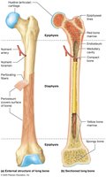
Long bones have a distinct structure that includes the diaphysis, epiphyses, articular cartilage, periosteum, medullary cavity, and endosteum.
Diaphysis: Shaft of the bone.
Epiphysis: Ends of the bone.
Articular Cartilage: Covers epiphysis, reduces friction at joints.
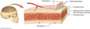
Periosteum: Dense irregular connective tissue membrane surrounding the bone.
Medullary Cavity: Hollow cavity within diaphysis containing bone marrow.
Endosteum: Lines inner surface of bone, contains bone cells.
Perforating Fibers: Anchor periosteum to underlying bone.
Structure of Short, Irregular, and Flat Bones
Bone Marrow Distribution
These bones contain bone marrow but lack a distinct marrow cavity. Their structure is adapted to their function and location in the body.
Bone Tissue Composition
Inorganic and Organic Components
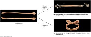
Bone tissue is composed of both inorganic and organic matrices, each contributing to bone strength and flexibility.
Inorganic Matrix: Makes up 65% of bone; mainly hydroxyapatite (calcium phosphate); provides hardness and resistance to compression.
Organic Matrix: Makes up 35% of bone; includes collagen fibers, proteoglycans, glycoproteins; provides flexibility and tensile strength.
Types of Bone Tissue
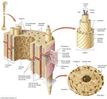
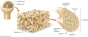
Compact and Spongy Bone
Bone tissue exists in two forms: compact bone and spongy bone, each with unique structural and functional properties.
Compact Bone: External layer; solid; resists stress; functional unit is the osteon (Haversian system).
Spongy Bone: Internal to compact bone; resists forces from multiple directions; forms a framework for bone marrow; contains trabeculae.
Bone Cells
Types and Functions
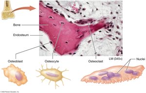
Bone growth and maintenance are regulated by four main cell types: osteoprogenitor cells, osteoblasts, osteocytes, and osteoclasts.
Osteoprogenitor (Osteogenic) Cells: Stem cells that differentiate into osteoblasts.
Osteoblasts: Bone-forming cells; secrete bone matrix.
Osteocytes: Mature bone cells; maintain bone structure and density.
Osteoclasts: Multinucleated cells; break down bone matrix for remodeling and calcium release.
Ossification (Bone Formation)
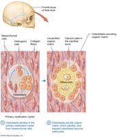
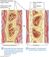
Intramembranous Ossification
Intramembranous ossification forms bones of the skull, facial bones, and clavicle. It involves the differentiation of mesenchymal cells into osteoblasts, secretion of organic matrix, and calcification.
Mesenchymal cells → osteogenic cells → osteoblasts
Osteoblasts secrete matrix, which calcifies
Trabeculae of early spongy bone form
Periosteum develops; osteoblasts lay down compact bone
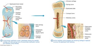
Endochondral Ossification
Endochondral ossification forms most bones, starting with a hyaline cartilage model. Chondroblasts differentiate into osteoblasts, and ossification begins at primary and secondary centers.
Hyaline cartilage model forms
Chondroblasts → osteoblasts
Primary ossification center develops in diaphysis
Secondary ossification centers develop in epiphyses

Growth of Bone
Appositional and Interstitial Growth
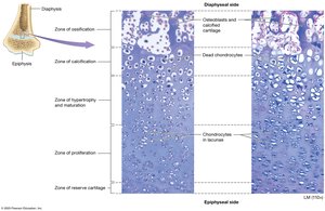
Bones grow in width (appositional growth) and length (interstitial growth). Interstitial growth occurs at the epiphyseal plate, where chondrocytes divide and new matrix is secreted.
Appositional Growth: Increases bone thickness.
Interstitial Growth: Lengthens bones at the epiphyseal plate.
Factors Affecting Bone Growth
Genetics, Hormones, and Nutrition
Bone growth is influenced by genetic factors, hormones, and nutrition. Essential minerals and vitamins are required for proper bone development and maintenance.
Minerals: Calcium, phosphorus, manganese, magnesium, fluoride.
Vitamins: D (calcium absorption), A (osteoblast activity), C (collagen synthesis), K and B12 (protein synthesis).
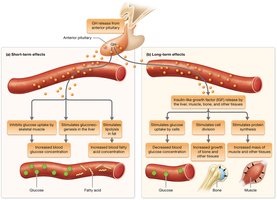
Hormones: Growth hormone, thyroid hormones, insulin, sex hormones.
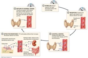
Hormonal Control of Calcium Homeostasis
Parathyroid Hormone and Calcitonin
Calcium levels in the blood are regulated by parathyroid hormone (PTH) and calcitonin. PTH increases blood calcium by stimulating osteoclasts, while calcitonin lowers blood calcium temporarily.
PTH: Released when blood calcium is low; increases osteoclast activity.
Calcitonin: Released by thyroid; lowers blood calcium in high doses.
Bone Remodeling
Continuous Process of Formation and Resorption
Bone remodeling is a lifelong process involving bone deposition by osteoblasts and resorption by osteoclasts. It maintains calcium homeostasis, repairs bone, and adapts to mechanical stress.
Bone Deposition: Formation of new bone.
Bone Resorption: Removal of old bone.
Mechanical Stress: Weight-bearing activities increase bone mass; lack of stress decreases bone mass.
Effects of Aging on Bone Tissue
Loss of Bone Mass and Brittleness
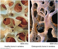
Aging leads to decreased sex hormones, resulting in loss of bone mass and increased brittleness. Osteoporosis is more common in older women due to greater bone resorption.
Loss of Bone Mass: Due to decreased calcium in bone matrix.
Brittleness: Due to decreased collagen production.
Disorders of Bones
Common Bone Disorders
Several disorders affect bone health, including osteoporosis, osteomalacia, rickets, Paget’s disease, and osteosarcoma.
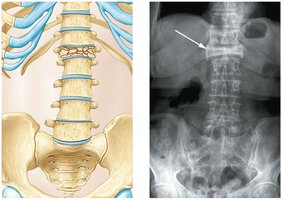
Osteoporosis: Low bone mass; bone resorption outpaces deposition.
Osteomalacia: Inadequate mineralization in adults.
Rickets: Insufficient vitamin D in children.
Paget’s Disease: Excessive bone deposition.
Osteosarcoma: Bone cancer.
Fracture and Repair of Bone
Types of Fractures
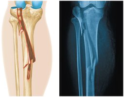
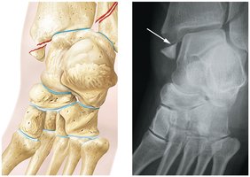
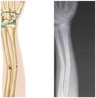
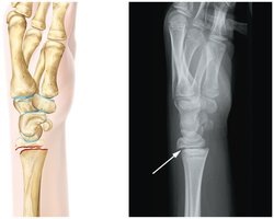
Bone fractures are classified by their characteristics and location. Common types include open, closed, comminuted, greenstick, impacted, Pott’s, Colles’, stress, and avulsion fractures.
Open (Compound): Bone protrudes through skin.
Closed (Simple): Bone does not break skin.
Comminuted: Bone is splintered or crushed.
Greenstick: Partial fracture; one side breaks, other bends.
Impacted: One end driven into another.
Pott’s: Fracture of fibula with tibial injury.
Colles’: Fracture of radius with distal fragment displacement.
Stress: Microscopic fissures.
Avulsion: Small piece of bone detached.
Healing of Bone Fractures
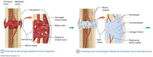
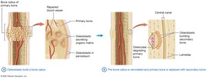
Fracture healing involves four main steps: hematoma formation, soft callus formation, bone callus formation, and remodeling.
Hematoma fills gap between bone fragments.
Fibroblasts and chondroblasts form soft callus.
Osteoblasts build bone callus.
Bone callus is remodeled and replaced with secondary bone.
Bone Density and Lifestyle Factors
Impact of Diet, Exercise, and Sunlight
Bone density is influenced by nutrition, physical activity, and exposure to sunlight (vitamin D synthesis). Poor diet, lack of exercise, and limited sunlight can lead to decreased bone density and increased fracture risk.
Calcium and Vitamin D: Essential for bone health.
Exercise: Stimulates bone formation.
Sunlight: Promotes vitamin D synthesis.
Additional info: Bone density is assessed by radiographs and blood calcium levels; lifestyle modifications can improve bone health and reduce fracture risk.
