 Back
CH 6: The Skeletal System: Structure, Function, and Physiology
Study Guide - Smart Notes
 Tailored notes based on your materials, expanded with key definitions, examples, and context.
Ch 6: The Skeletal System: Structure, Function, and Physiology
Major Functions of the Skeletal System
The skeletal system is a dynamic organ system that provides the framework for the body and supports numerous physiological processes. It consists of bones, cartilages, ligaments, and other connective tissues.
Support: Provides structural support and a framework for the attachment of soft tissues and organs.
Storage of Minerals and Lipids: Bones store minerals such as calcium and phosphate, essential for physiological processes, and lipids in yellow bone marrow for energy reserves.
Blood Cell Production: Red and white blood cells, as well as platelets, are produced in red bone marrow.
Protection: Skeletal structures protect vital organs (e.g., skull protects the brain, ribs protect the heart and lungs).
Leverage: Bones act as levers for muscle action, enabling movement.
Classification and Structure of Bones
Bone Shapes
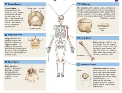
Bones are classified by their shapes, which relate to their functions and locations in the body.
Sutural Bones: Small, flat, irregular bones found between the flat bones of the skull.
Irregular Bones: Complex shapes, such as vertebrae.
Short Bones: Boxy, with nearly equal dimensions (e.g., carpal bones).
Flat Bones: Thin, parallel surfaces (e.g., parietal bone of the skull, sternum).
Long Bones: Long and slender, with a shaft and two ends (e.g., femur, humerus).
Sesamoid Bones: Small, round, and flat; develop within tendons (e.g., patella).
Bone Markings
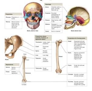
Bone markings are surface features that serve as attachment sites for muscles, tendons, and ligaments, or as passages for nerves and blood vessels. They include projections, depressions, and openings.
Projections: Sites of muscle and ligament attachment (e.g., tuberosity, crest).
Openings: Allow passage of blood vessels and nerves (e.g., foramen, canal).
Depressions: Accommodate other structures (e.g., fossa, groove).
Bone Structure
Bones have distinct structural regions:
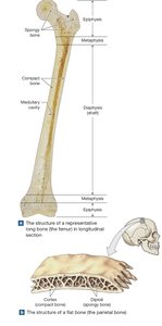
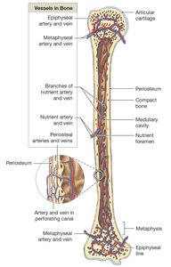
Diaphysis: The shaft, composed of compact bone surrounding the medullary cavity.
Epiphysis: Expanded ends, mostly spongy bone covered by compact bone.
Metaphysis: The narrow region connecting diaphysis and epiphysis.
Medullary Cavity: Central cavity containing bone marrow.
Flat Bones: Consist of two layers of compact bone with spongy bone (diploë) in between.
Bone Matrix and Cellular Composition
Bone Matrix
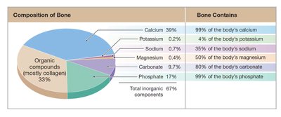
The bone matrix is composed of inorganic and organic components:
Inorganic: Calcium phosphate (Ca3(PO4)2) forms hydroxyapatite crystals, providing compressive strength.
Organic: Collagen fibers provide flexibility and tensile strength.
The combination of collagen and hydroxyapatite gives bone its unique properties: strong, somewhat flexible, and resistant to shattering.
Bone Cells
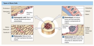
There are four main types of bone cells, each with distinct functions:
Cell Type | Function |
|---|---|
Osteogenic cells | Stem cells that differentiate into osteoblasts; important for bone repair. |
Osteoblasts | Produce new bone matrix (osteoid) and initiate ossification. |
Osteocytes | Mature bone cells that maintain the bone matrix and participate in repair. |
Osteoclasts | Multinucleate cells that resorb bone matrix, releasing minerals into the blood. |
Compact and Spongy Bone Structure
Compact Bone
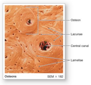
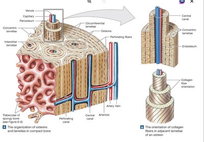
Compact bone is organized into osteons (Haversian systems), which are cylindrical structures aligned parallel to the long axis of the bone. Each osteon consists of concentric lamellae surrounding a central canal containing blood vessels and nerves.
Lamellae: Layers of bone matrix.
Lacunae: Small spaces housing osteocytes.
Canaliculi: Tiny channels for nutrient and waste exchange.
Perforating (Volkmann's) Canals: Connect central canals transversely.
Spongy Bone
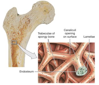
Spongy bone (trabecular bone) consists of a network of trabeculae, which are oriented along lines of stress. It is lighter than compact bone and contains spaces filled with bone marrow.
Trabeculae: Supporting bundles of bony fibers.
Red Bone Marrow: Site of hematopoiesis (blood cell formation).
Yellow Bone Marrow: Stores fat for energy.
Functional Integration
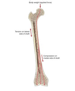
Compact bone provides strength for weight-bearing, while spongy bone reduces skeletal weight and supports marrow. The distribution of forces across the femur demonstrates how these structures work together to resist compression and tension.
Surface Coverings of Bone
Periosteum
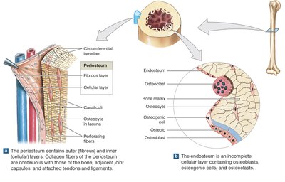
The periosteum is a membrane covering the outer surface of bones, except at joints. It consists of an outer fibrous layer and an inner cellular layer. Functions include isolating bone, providing a route for blood vessels and nerves, and participating in growth and repair.
Endosteum
The endosteum is an incomplete cellular layer lining the medullary cavity and covering trabeculae. It contains osteogenic cells, osteoblasts, and osteoclasts, and is active in bone growth, repair, and remodeling.
Bone Formation and Growth
Ossification
Ossification is the process of bone formation, occurring via two mechanisms:
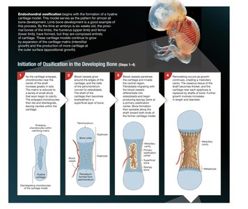
Endochondral Ossification: Bone replaces a cartilage model; primary mechanism for long bone development.
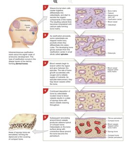
Intramembranous Ossification: Bone develops directly from mesenchymal tissue; forms flat bones of the skull, mandible, and clavicles.
Bone Growth
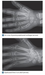
Interstitial Growth: Increases bone length via growth at the epiphyseal plate.
Appositional Growth: Increases bone diameter by adding layers to the outer surface.
Bone Remodeling and Homeostasis
Bone Remodeling
Bone remodeling is a continuous process involving osteoblasts, osteoclasts, and osteocytes. It replaces old bone matrix with new, adapts bone structure to stress, and regulates mineral homeostasis.
High Turnover: About 20% of adult bone is replaced annually.
Imbalance: Excess osteoclast activity weakens bone; excess osteoblast activity strengthens bone.
Factors Affecting Bone Development
Exercise
Physical activity stimulates bone formation and increases bone density. Inactivity leads to bone loss and increased risk of fractures.
Nutrition and Hormones
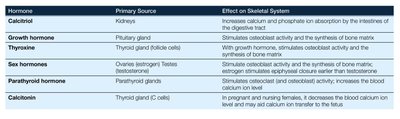
Bone health depends on adequate intake of minerals (calcium, phosphorus, magnesium), vitamins (D, C, A, K, B12), and hormones (growth hormone, thyroxine, sex hormones, parathyroid hormone, calcitonin).
Hormone | Primary Source | Effect on Skeletal System |
|---|---|---|
Calcitriol | Kidneys | Increases calcium and phosphate absorption in intestines |
Growth hormone | Pituitary gland | Stimulates osteoblast activity and bone matrix synthesis |
Thyroxine | Thyroid gland | Stimulates cell metabolism and osteoblast activity |
Sex hormones | Ovaries/Testes | Stimulate osteoblast activity and bone growth |
Parathyroid hormone | Parathyroid glands | Stimulates osteoclast activity; increases blood calcium |
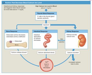
Calcitonin | Thyroid gland (C cells) | Decreases blood calcium; may aid calcium transfer to fetus |
Calcium Homeostasis and Bone Physiology
Calcium as a Mineral Reserve
Bone contains 99% of the body's calcium and serves as a reservoir for maintaining blood calcium levels. Calcium is essential for nerve impulse transmission, muscle contraction, and blood clotting.
Hormonal Regulation of Calcium
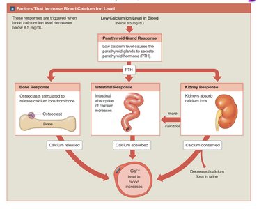
Calcium homeostasis is regulated by parathyroid hormone (PTH) and calcitonin:
PTH: Increases blood calcium by stimulating osteoclasts, increasing intestinal absorption (via calcitriol), and reducing renal excretion.
Calcitonin: Decreases blood calcium by inhibiting osteoclasts, reducing intestinal absorption, and increasing renal excretion.
Clinical Correlations
Pituitary Growth Failure: Inadequate growth hormone leads to short stature.
Gigantism/Acromegaly: Excess growth hormone causes abnormal bone growth.
Marfan Syndrome: Genetic disorder affecting connective tissue, resulting in long limbs and increased risk of cardiovascular issues.
Osteomalacia/Rickets: Poor bone mineralization due to vitamin D deficiency, leading to soft, weak bones.
Additional info: The skeletal system is highly adaptive, responding to mechanical stress, nutritional status, and hormonal signals. Its health is critical for overall physiological function and longevity.
