 Back
BackThe Skeletal System: Structure, Function, and Disorders
Study Guide - Smart Notes
 Tailored notes based on your materials, expanded with key definitions, examples, and context.
The Skeletal System
Introduction to the Skeletal System
The skeletal system is a fundamental organ system in the human body, consisting of approximately 206 bones in adults. It also includes cartilages, ligaments, joints, and other connective tissues that stabilize or connect bones.
Protection: Shields vital organs (e.g., skull protects the brain).
Support: Provides structural framework for the body.
Blood cell production: Hematopoiesis occurs in bone marrow.
Storage of lipids and minerals: Stores adipose tissue, calcium, and phosphate.
Leverage: Muscles attach to bones for movement.
Bone Connective Tissue Composition
Bone tissue is a specialized connective tissue composed of collagen fibers and a calcified matrix.
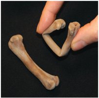
Collagen: Makes up one-third of bone weight; provides strength and flexibility.
Calcified matrix (calcium phosphate): Makes up two-thirds of bone weight; provides hardness and resistance to shattering.
Combination: Collagen and calcified crystals together make bone strong, flexible, and highly resistant to shattering.
Axial and Appendicular Skeleton
The skeleton is divided into two main regions:
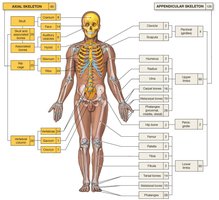
Axial skeleton: Includes bones of the skull, thorax, and vertebral column; forms the longitudinal axis of the body.
Appendicular skeleton: Includes bones of the limbs and pectoral/pelvic girdles; attaches limbs to the axial skeleton.
Bone Classifications
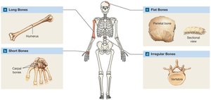
Bones are classified based on their shape, which is closely related to their function.
Flat bones: Protect internal organs (e.g., skull, ribs).
Long bones: Support weight and facilitate movement (e.g., femur, humerus).
Short bones: Provide stability and some movement (e.g., carpals).
Irregular bones: Complex shapes (e.g., vertebrae).
Sesamoid bones: Embedded in tendons (e.g., patella).
Structure of a Long Bone
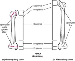
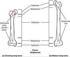
Long bones have a distinct structure, including the diaphysis (shaft), epiphyses (ends), metaphysis, and epiphyseal plate/line.
Diaphysis: Shaft of the bone.
Epiphysis: Ends of the bone.
Metaphysis: Region between diaphysis and epiphysis.
Epiphyseal plate/line: Growth plate in children, becomes epiphyseal line in adults.
Types of Bone Tissue
Bone tissue is categorized into two main types:
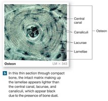
Compact bone: Covers the outside of bone surfaces; composed of osteons; resists stress in a specific direction.
Spongy bone: Found inside bone; reduces bone weight and provides strength; consists of trabeculae with spaces filled with marrow.
Bone Cells
Both compact and spongy bone contain three main typecs of cells:
Osteocytes: Mature bone cells that maintain the matrix.
Osteoblasts: Cells that build new bone matrix.
Osteoclasts: Cells that break down bone matrix.
Bone Development (Ossification)
Bone formation begins with cartilaginous models in embryos and continues into adulthood.
Intramembranous ossification: Bone develops within a membrane.
Endochondral ossification: Bone replaces a cartilage model.
Types of Bone Growth
Bone grows in two ways:
Interstitial growth: Growth in length at the epiphyseal plate; closure results in the epiphyseal line.
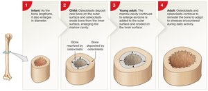
Appositional growth: Growth in diameter/thickness.
Bone Remodeling and Homeostasis
Bone tissue is constantly remodeled, with 3-5% of bone calcium exchanged each year.
Osteocytes: Maintain matrix.
Osteoclasts: Remove matrix.
Osteoblasts: Build matrix.
Factors: Nutrition (Vitamins A, C, D), exercise, hormones (GH, thyroid, sex hormones, calcitonin, PTH).
Hormonal Regulation of Calcium Homeostasis
Calcitonin: Promotes calcium deposit in bone, decreases blood calcium, inhibits osteoclasts.
Parathyroid hormone (PTH): Increases blood calcium, stimulates osteoclasts.
Skeletal Disorders
Atypical Skeletal Growth
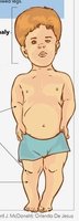
Achondroplasia: Slow growth of epiphyseal cartilage; short, stocky limbs.
Gigantism: Overproduction of growth hormone before puberty; delayed puberty, excessive growth.
Acromegaly: Overproduction of growth hormone after epiphyseal closure; bones thicken, especially in face, jaw, hands.
Osteopenia and Osteoporosis
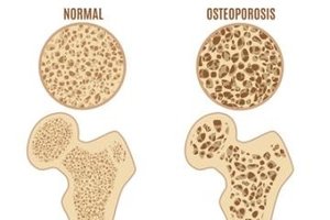
Osteopenia: Age-related loss of cartilage from joints.
Osteoporosis: Disease resulting in loss of bone density and mass; bones become porous and fragile.
Bone Fractures
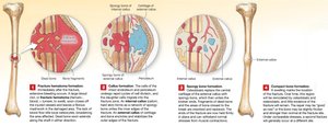
Fracture: Crack or break in bone; classified as closed (simple) or open (compound).
Joints
Joints are classified by degree of movement and structure.
Synarthrotic: No movement (e.g., sutures in skull).
Amphiarthrotic: Little movement (e.g., symphysis between pubic bones).
Diarthrotic: Free movement (e.g., synovial joints).
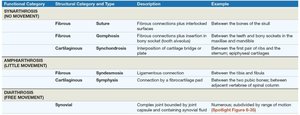
Functional Category | Structural Category and Type | Description | Example |
|---|---|---|---|
Synarthrosis (No Movement) | Fibrous: Suture | Fibrous connections plus interlocked surfaces | Between bones of the skull |
Synarthrosis (No Movement) | Fibrous: Gomphosis | Fibrous connection plus insertion in bone socket | Between teeth and bony sockets in the maxilla and mandible |
Synarthrosis (No Movement) | Cartilaginous: Synchondrosis | Interposition of cartilage bridge or plate | Between first pair of ribs and the sternum; epiphyseal cartilage |
Amphiarthrosis (Little Movement) | Fibrous: Syndesmosis | Ligamentous connection | Between the tibia and fibula |
Amphiarthrosis (Little Movement) | Cartilaginous: Symphysis | Connection by fibrocartilage pad | Between adjacent vertebrae of spinal column |
Diarthrosis (Free Movement) | Synovial | Complex joint bounded by joint capsule and containing synovial fluid | Numerous, subdivided by range of motion |
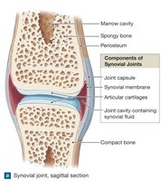
Synovial Joints
Synovial joints are the most complex and allow free movement.
Articular cartilage: Reduces friction and absorbs shock.
Synovial fluid: Lubricates joint, nourishes cartilage, removes waste.
Types and Examples of Synovial Joints
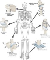
Synovial joints are classified by their shape and movement:
Ball-and-socket: Shoulder, hip.
Hinge: Elbow, knee.
Pivot: Atlas/axis vertebrae.
Gliding: Wrist, ankle.
Saddle: Thumb.
Condyloid: Fingers.
Skeletal Disorders of Joints: Arthritis
Arthritis: Inflamed, swollen joints; includes over 100 types of inflammatory or degenerative diseases.
Rheumatoid arthritis: Chronic inflammatory disease; autoimmune disorder where the body attacks synovial joint membrane.
Summary Table: Skeletal System Functions and Disorders
Function/Disorder | Description |
|---|---|
Protection | Shields vital organs |
Support | Framework for body |
Blood cell production | Hematopoiesis in marrow |
Storage | Calcium, phosphate, lipids |
Leverage | Muscle attachment for movement |
Achondroplasia | Slow growth of epiphyseal cartilage |
Gigantism | Excess GH before puberty |
Acromegaly | Excess GH after puberty |
Osteopenia | Age-related cartilage loss |
Osteoporosis | Loss of bone density |
Arthritis | Inflamed joints |
Rheumatoid arthritis | Autoimmune attack on joints |
Key Equations
Calcium Homeostasis:
Additional info:
Vitamin D is essential for calcium absorption and bone health.
Exercise increases bone density and strength by stimulating osteoblast activity.
