 Back
The Special Senses: Structure of the Eye and Ear
Study Guide - Smart Notes
 Tailored notes based on your materials, expanded with key definitions, examples, and context.
The Special Senses
Structure of the Eyeball
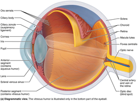
The human eyeball is a complex organ responsible for vision. Its wall consists of three main layers, and its internal cavity is filled with fluids called humors. The lens divides the internal cavity into anterior and posterior segments.
Fibrous Layer: The outermost layer, composed of dense avascular connective tissue. It includes the sclera (opaque, protective, and shapes the eyeball) and the cornea (transparent, allows light entry, bends light, and contains pain receptors).
Vascular Layer: The middle layer, not detailed here but includes structures such as the choroid, ciliary body, and iris.
Inner Layer (Retina): Originates as an outpocketing of the brain and contains millions of photoreceptor cells, neurons, and glial cells. It is a delicate two-layered membrane: the outer pigmented layer and the inner neural layer.
Internal Cavity: Filled with humors (aqueous and vitreous) that help maintain the shape and optical properties of the eye.
Lens: A biconvex structure that focuses light onto the retina.
Microscopic Anatomy of the Retina
The retina is composed of two main layers:
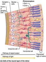
Pigmented Layer: A single-cell-thick lining next to the choroid, extending anteriorly to cover the ciliary body and iris. It absorbs light, prevents scattering, phagocytizes photoreceptor fragments, and stores vitamin A.
Neural Layer: Transparent, running anteriorly to the margin of the ciliary body. It contains three main types of neurons: photoreceptors (rods and cones), bipolar cells, and ganglion cells. Signals spread from photoreceptors to bipolar cells to ganglion cells, whose axons exit the eye as the optic nerve.
Photoreceptors: Rods and Cones
The retina contains approximately a quarter-billion photoreceptors, which are classified as rods and cones:
Rods: Specialized for dim light and peripheral vision. They are more numerous and sensitive to light but do not provide color vision or sharp images. Most abundant at the periphery of the retina.
Cones: Responsible for bright light and high-resolution color vision. Concentrated in the macula lutea, especially in the fovea centralis, which provides the best visual acuity.
Blood Supply to the Retina
The retina receives blood from two sources:
Choroid: Supplies the outer third, including photoreceptors.
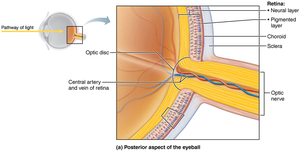
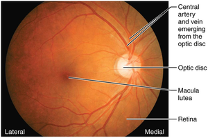
Central Artery and Vein of the Retina: Supply the inner two-thirds and enter/exit the eye at the center of the optic nerve. These vessels are visible in a living person using an ophthalmoscope.
Structure of the Ear
Major Regions of the Ear
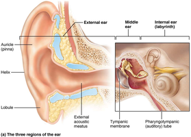
The ear is divided into three major areas, each with distinct functions:
External (Outer) Ear: Involved in hearing only. Consists of the auricle (pinna), which funnels sound waves into the auditory canal, and the external acoustic meatus (auditory canal), which transmits sound waves to the tympanic membrane (eardrum).
Middle Ear (Tympanic Cavity): Also involved in hearing only. Contains the auditory ossicles (malleus, incus, stapes) that transmit vibratory motion from the eardrum to the oval window. The pharyngotympanic (auditory) tube connects the middle ear to the nasopharynx and equalizes pressure.
Internal (Inner) Ear: Responsible for both hearing and equilibrium. Contains the vestibule, semicircular canals, and cochlea, each with specialized receptor regions.
Summary Table: Internal Ear
The internal ear consists of bony and membranous labyrinths, each with specific functions and receptor regions:
BONY LABYRINTH | MEMBRANOUS LABYRINTH | FUNCTION | RECEPTOR REGION |
|---|---|---|---|
Semicircular canals | Semicircular ducts | Equilibrium: rotational (angular) acceleration | Crista ampullaris |
Vestibule | Utricle and saccule | Equilibrium: head position relative to gravity, linear acceleration | Macula |
Cochlea | Cochlear duct (scala media) | Hearing | Spiral organ |

