 Back
BackThe Spinal Cord and Peripheral Nervous System: Structure, Function, and Autonomic Divisions
Study Guide - Smart Notes
 Tailored notes based on your materials, expanded with key definitions, examples, and context.
IV. The Spinal Cord
a. Function
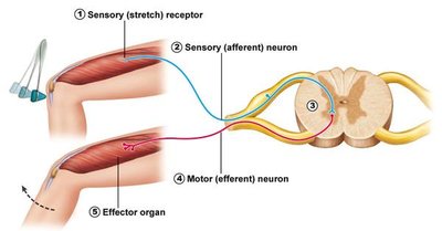
The spinal cord serves as a critical communication pathway between the peripheral nervous system (PNS) and the brain, facilitating both sensory and motor information transfer. It is also a major reflex center, responsible for executing rapid, involuntary responses to stimuli, such as the knee-jerk reflex.
Conduction Pathways: Ascending tracts carry sensory information to the brain, while descending tracts transmit motor commands from the brain to the body.
Reflex Center: The spinal cord mediates reflexes, which are automatic responses to specific stimuli, bypassing the brain for faster reaction times.
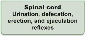
Examples of Reflexes: Knee-jerk, urination, defecation, erection, and ejaculation reflexes.
b. Anatomy
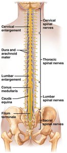
The spinal cord is a cylindrical structure located within the vertebral canal, extending from the foramen magnum to the level of the first or second lumbar vertebra. It is protected by vertebrae, meninges, and cerebrospinal fluid (CSF).
Gray Matter: Located centrally, shaped like a butterfly or the letter 'H', and contains neuron cell bodies and interneurons.
White Matter: Surrounds the gray matter and consists of myelinated axons organized into columns (funiculi) that carry information up and down the spinal cord.
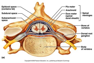
Meninges: Three connective tissue layers (dura mater, arachnoid mater, pia mater) that protect the spinal cord and contain CSF in the subarachnoid space.
1. Gray Matter
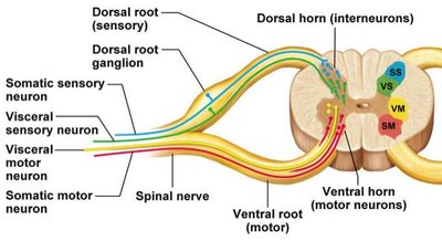
The gray matter of the spinal cord is divided into dorsal (posterior), ventral (anterior), and lateral horns, each with distinct functions:
Dorsal Horns: Contain interneurons and axon terminals of sensory neurons (somatic and visceral).
Ventral Horns: House cell bodies of somatic motor neurons and interneurons; send signals to skeletal muscles.
Lateral Horns: Present in thoracic and upper lumbar regions; contain visceral motor neuron cell bodies for autonomic (sympathetic) output.
2. White Matter
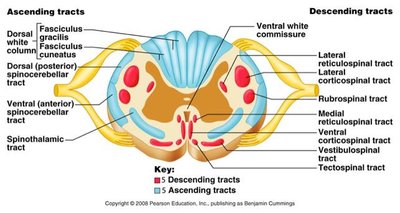
White matter consists of myelinated axons organized into dorsal, lateral, and ventral columns (funiculi). These columns contain ascending (sensory) and descending (motor) tracts that connect the spinal cord with the brain.
Ascending Tracts: Carry sensory information to the brain.
Descending Tracts: Transmit motor commands from the brain to the body.
3. Meninges
The spinal cord is surrounded by three protective membranes:
Dura Mater: Outermost, tough layer.
Arachnoid Mater: Middle, web-like layer.
Pia Mater: Innermost, delicate layer adhering to the spinal cord surface.
Spaces: Epidural (fat), subdural, and subarachnoid (contains CSF).
Clinical Note: A spinal tap (lumbar puncture) samples CSF from the subarachnoid space.
V. Peripheral Nervous System (PNS)
a. Introduction
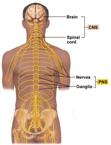
The peripheral nervous system (PNS) consists of nerves and ganglia outside the brain and spinal cord. It connects the CNS to limbs and organs, facilitating communication throughout the body.
Nerves: Bundles of axons (nerve fibers) that transmit signals between the CNS and the body.
Ganglia: Collections of neuron cell bodies located outside the CNS.
Most nerves are mixed, carrying both sensory (afferent) and motor (efferent) fibers; cranial nerves are exceptions.
b. Spinal Nerves & Associated Structures
Spinal nerves are short, mixed nerves that emerge from the spinal cord and branch into dorsal and ventral rami. They connect the CNS to sensory receptors, muscles, and glands.
Dorsal Root: Contains sensory (afferent) fibers; has a dorsal root ganglion with sensory neuron cell bodies.
Ventral Root: Contains motor (efferent) fibers from motor neurons in the ventral horn.
Dorsal Ramus: Innervates muscles and skin of the back.
Ventral Ramus: Innervates the rest of the trunk and limbs.
c. Autonomic Nervous System (ANS)
The autonomic nervous system (ANS) is a division of the PNS that controls involuntary functions by regulating smooth muscle, cardiac muscle, and glands. It is subdivided into the sympathetic and parasympathetic nervous systems.
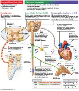
Somatic Motor System: Controls voluntary movements of skeletal muscle; one neuron from CNS to effector.
Visceral Motor (Autonomic) System: Controls involuntary effectors; two-neuron chain (preganglionic and postganglionic).
1. Introduction
Somatic Motor: One synapse, voluntary, targets skeletal muscle.
Autonomic (Visceral Motor): Two synapses, involuntary, targets smooth muscle, cardiac muscle, and glands.
2. Sympathetic Nervous System
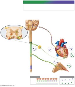
The sympathetic nervous system prepares the body for 'fight or flight' responses. It arises from thoracic and lumbar spinal nerves.
General Features: Preganglionic fibers are short; ganglia are near the spinal cord; postganglionic fibers are long and highly branched.
Neurotransmitters: Preganglionic neurons release acetylcholine (ACh); postganglionic neurons release norepinephrine (NE).
Physiological Responses: Increased heart rate, bronchiole dilation, decreased digestion, increased blood flow to muscles, glucose release, and sweating.
Adrenal Medulla: Acts as a modified sympathetic ganglion, releasing epinephrine (adrenaline) and norepinephrine into the bloodstream.
3. Parasympathetic Nervous System
The parasympathetic nervous system supports 'rest and digest' activities. It arises from cranial nerves (especially the vagus nerve) and sacral spinal nerves.
General Features: Preganglionic fibers are long; ganglia are near or within effector organs; postganglionic fibers are short and have few branches.
Neurotransmitters: Both pre- and postganglionic neurons release acetylcholine (ACh).
Physiological Responses: Decreased heart rate, increased gastrointestinal motility and secretions, bronchiole constriction, and organ-specific control.
d. Sensory System
The sensory system detects and transduces environmental stimuli into electrical signals, which are then transmitted to the CNS for processing.
Sensory Receptors: Specialized cells or nerve endings that respond to specific types of stimuli.
Types of Sensory Receptors:
Mechanoreceptors: Detect touch, pressure, vibration, and stretch.
Thermoreceptors: Detect temperature changes.
Nociceptors: Detect pain.
Chemoreceptors: Detect chemical stimuli (e.g., taste, smell).
Photoreceptors: Detect light (vision).
General Senses: Widely distributed receptors for somatic (body wall, limbs) and visceral (internal organs) sensations.
Special Senses: Localized, complex sense organs for vision, hearing, equilibrium, taste, and smell.
