 Back
BackThe Urinary System: Structure, Function, and Regulation
Study Guide - Smart Notes
 Tailored notes based on your materials, expanded with key definitions, examples, and context.
The Urinary System
Overview
The urinary system is essential for filtering blood, removing waste, and maintaining the body's fluid, electrolyte, and acid-base balance. It consists of the kidneys, ureters, urinary bladder, and urethra. The kidneys are the primary organs responsible for urine formation and homeostatic regulation.
Location and External Anatomy of the Kidneys
Position and Structure
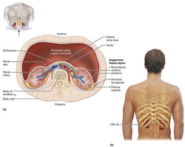
Retroperitoneal Position: The kidneys are located behind the peritoneum in the superior lumbar region, extending from the 12th thoracic to the 3rd lumbar vertebrae.
Asymmetry: The right kidney is slightly lower than the left due to the presence of the liver.
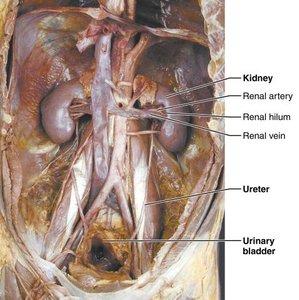
Surfaces: The lateral surface is convex, and the medial surface is concave, featuring the renal hilus—a vertical cleft leading to the renal sinus.
Entry/Exit Points: Ureters, renal blood vessels, lymphatics, and nerves enter and exit at the hilus.
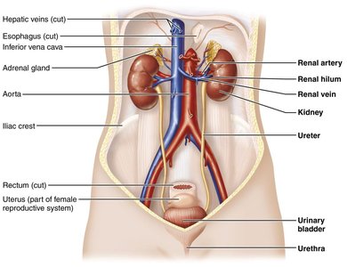
Organs of the Urinary System
Major Components
Kidneys: Filter 200–400 liters of fluid from the blood daily, removing toxins, metabolic wastes, and excess ions in urine. They regulate blood volume and composition, produce renin (for blood pressure regulation), erythropoietin (stimulates RBC production), and activate vitamin D.
Ureters: Paired tubes that transport urine from the kidneys to the bladder.
Urinary Bladder: Temporary storage reservoir for urine.
Urethra: Transports urine from the bladder out of the body.
Internal Anatomy of the Kidneys
Regions and Structures
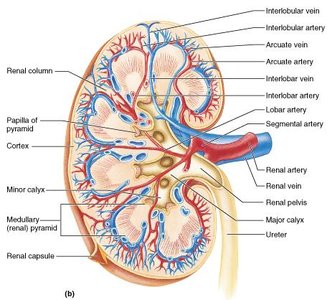
Cortex: The outer, light-colored, granular region.
Medulla: Contains cone-shaped renal pyramids made of parallel bundles of urine-collecting tubules.
Renal Columns: Inward extensions of cortical tissue separating the pyramids.
Renal Lobe: Consists of a medullary pyramid and its surrounding cortical tissue.
Renal Pelvis: Funnel-shaped tube within the renal sinus that collects urine from the major calyces.
Major and Minor Calyces: Collect urine from the papillae and empty it into the renal pelvis.

Blood and Nerve Supply
Vascularization and Innervation
About one-fourth (1200 mL) of systemic cardiac output flows through the kidneys each minute.
Arterial and venous blood flow follow similar paths through the kidney.
The renal plexus provides nerve supply to the kidneys.
The Nephron: Functional Unit of the Kidney
Structure and Types
Nephron: The functional unit responsible for urine formation, consisting of the renal corpuscle and renal tubule.
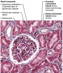
Renal Corpuscle: Includes the glomerulus (a tuft of capillaries) and the glomerular (Bowman’s) capsule (surrounds the glomerulus).
Glomerular Endothelium: Fenestrated epithelium allowing solute-rich, protein-free filtrate to pass into the capsule.
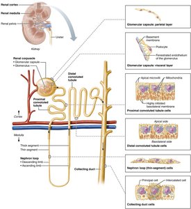
Renal Tubule Segments
Proximal Convoluted Tubule (PCT): Cuboidal cells with microvilli; reabsorbs water and solutes, secretes substances.
Nephron Loop (Loop of Henle): Descending limb (thin, simple squamous cells, permeable to water), ascending limb (thick, cuboidal/columnar cells, permeable to solutes).
Distal Convoluted Tubule (DCT): Cuboidal cells, mainly involved in secretion.
Collecting Ducts: Contain intercalated cells (acid-base balance) and principal cells (water/salt balance, responsive to ADH and aldosterone).
Types of Nephrons
Cortical Nephrons: 85% of nephrons, located in the cortex, short nephron loops.
Juxtamedullary Nephrons: Located at the cortex-medulla junction, long loops deeply invade the medulla, essential for producing concentrated urine.
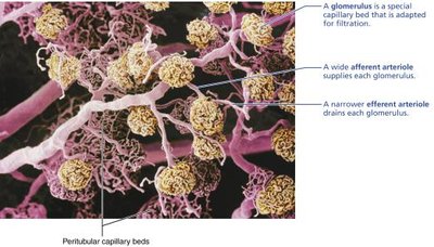
Capillary Beds of the Nephron
Glomerulus and Peritubular Capillaries
Each nephron has two capillary beds: the glomerulus (filtration) and peritubular capillaries (reabsorption).
Glomerulus is fed by an afferent arteriole and drained by an efferent arteriole.
Blood pressure in the glomerulus is high, promoting filtration; peritubular capillaries are low-pressure and adapted for absorption.
Vasa Recta: Long, straight capillaries associated with juxtamedullary nephrons, important for urine concentration.
Juxtaglomerular Apparatus (JGA)
Structure and Function
Located where the distal tubule contacts the afferent arteriole.
Juxtaglomerular (JG) Cells: Smooth muscle cells with secretory granules containing renin; act as mechanoreceptors.
Macula Densa: Tall, closely packed distal tubule cells; function as chemoreceptors/osmoreceptors (monitor NaCl concentration).
Mesangial Cells: Regulate glomerular filtration rate.
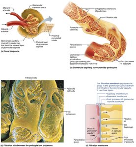
Mechanism of Urine Formation
Three Major Processes
Glomerular Filtration: Passive process where hydrostatic pressure forces fluids and solutes through a filtration membrane.
Tubular Reabsorption: Selective movement of substances from filtrate back into the blood.
Tubular Secretion: Reverse process of reabsorption; substances move from blood into filtrate for excretion.
Filtration Membrane
Composed of three layers: fenestrated endothelium, visceral membrane (podocytes), and fused basement membrane.
Allows passage of water and small solutes but not proteins or blood cells.
Net Filtration Pressure (NFP)
Responsible for filtrate formation.
Calculated as:
Where: = Glomerular hydrostatic pressure = Oncotic pressure of glomerular blood = Capsular hydrostatic pressure
Glomerular Filtration Rate (GFR)
Total amount of filtrate formed per minute by the kidneys.
Directly proportional to NFP, surface area, and membrane permeability.
Regulated by intrinsic (renal autoregulation) and extrinsic (neural, hormonal) mechanisms.
Tubular Reabsorption and Secretion
Reabsorption
Most filtrate is reabsorbed into the blood via transcellular or paracellular routes.
All organic nutrients are reabsorbed; water and ion reabsorption are hormonally regulated (ADH, aldosterone, ANP, PTH).
Sodium reabsorption is primarily active, driven by the Na+-K+ ATPase pump.
Secretion
Moves substances from blood into filtrate (e.g., drugs, excess K+, H+ for pH control).
Important for eliminating substances not filtered and controlling blood pH.
Regulation of Urine Concentration and Volume
Countercurrent Mechanism
Maintains the medullary osmotic gradient, allowing the kidneys to concentrate urine.
Countercurrent Multiplier: Nephron loops of juxtamedullary nephrons create the gradient.
Countercurrent Exchanger: Vasa recta preserve the gradient while delivering nutrients.
Hormonal Regulation
ADH: Increases water reabsorption in collecting ducts, producing concentrated urine.
Aldosterone: Increases Na+ reabsorption and K+ secretion.
ANP: Promotes Na+ excretion.
PTH: Increases Ca2+ reabsorption.
Renal Clearance
Definition and Equation
Volume of plasma cleared of a substance per minute (mL/min).
Used to determine GFR, detect glomerular damage, and monitor renal disease.
Where: = Renal clearance rate = Concentration of substance in urine (mg/mL) = Urine flow rate (mL/min) = Concentration in plasma (mg/mL)
Physical and Chemical Characteristics of Urine
Physical Properties
Color: Clear, pale to deep yellow (urochrome pigment).
Odor: Slightly aromatic; ammonia odor develops upon standing.
pH: Slightly acidic (pH 6), range 4.5–8.0.
Specific Gravity: 1.001–1.035, depending on solute concentration.
Chemical Composition
95% water, 5% solutes.
Nitrogenous wastes: Urea, uric acid, creatinine.
Other solutes: Na+, K+, phosphate, sulfate, Ca2+, Mg2+, HCO3-.
Ureters, Urinary Bladder, and Urethra
Ureters
Slender tubes conveying urine from kidneys to bladder.
Enter bladder at an angle to prevent backflow.
Wall: Transitional epithelium, smooth muscle, fibrous connective tissue.
Urinary Bladder
Muscular sac for temporary urine storage.
Located retroperitoneally on the pelvic floor.
Wall: Transitional epithelium, thick muscle, fibrous adventitia.
Trigone: Triangular area prone to infection.
Urethra
Muscular tube draining urine from bladder to exterior.
Internal urethral sphincter (involuntary) and external urethral sphincter (voluntary).
Female urethra: Short, anterior to vaginal opening.
Male urethra: Prostatic, membranous, and spongy regions.
