 Back
BackThe Urinary System: Structure, Function, and Physiology
Study Guide - Smart Notes
 Tailored notes based on your materials, expanded with key definitions, examples, and context.
The Urinary System
Overview and Objectives
The urinary system is essential for maintaining homeostasis by regulating the composition and volume of blood, removing waste products, and balancing electrolytes and acid-base levels. The following study notes cover the anatomy, functions, and physiological processes of the urinary system, including the role of nephrons in urine formation.
Learning Goal 1: Identify the organs and structures of the urinary system.
Learning Goal 2: Describe the functions performed by the urinary system.
Learning Goal 3: Explain the detailed functional anatomy of the organs of the urinary system.
Learning Goal 4: Describe nephron function in regulating blood chemistry and forming urine.
Learning Goal 5: Trace the route urine travels from formation to the body exterior.
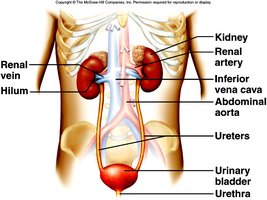
Organs of the Urinary System
The urinary system consists of several organs that work together to filter blood, remove waste, and excrete urine.
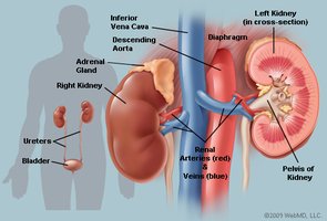
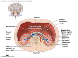
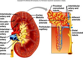
Kidneys: Major excretory organs located in the superior lumbar region, outside the parietal peritoneum. They filter blood and produce urine.
Ureters: Tubes that carry urine from the kidneys to the urinary bladder.
Urinary Bladder: Stores urine until it is expelled from the body.
Urethra: Conducts urine from the bladder to the exterior of the body.
Functions of the Urinary System
The urinary system performs several vital functions to maintain internal balance and health.
Homeostasis: Regulates the composition and volume of blood and internal fluids.
Waste Removal: Eliminates toxins, metabolic wastes, and excess ions via urine.
Blood Pressure Regulation: Produces renin, which helps maintain blood pressure.
Red Blood Cell Production: Produces erythropoietin, stimulating red blood cell formation.
Vitamin D Activation: Converts vitamin D to its active form, necessary for calcium absorption.
Gluconeogenesis: Generates glucose from non-carbohydrate sources during starvation or intense exercise.
Key Terminology
Renal: Refers to anything related to the kidneys.
Filtration: The process of filtering substances out of the blood to produce filtrate.
Reabsorption: Movement of substances from filtrate back into the bloodstream.
Secretion: Movement of substances from the blood into the renal tubule for excretion.
Nephrons: Structural and functional units of the kidneys responsible for urine formation.
Micturition: The process of emptying the bladder.
Electrolyte: Ions such as Na+, K+, Cl- that are important for cellular function.
Nitrogenous: Refers to waste products containing nitrogen, such as urea.
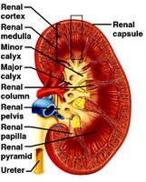
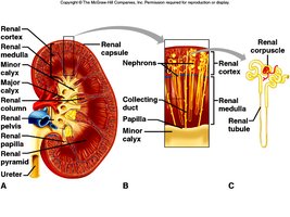
Functional Anatomy of the Urinary System
The kidneys have distinct external and internal anatomical features that support their function.
External Anatomy: Includes the renal capsule (innermost, prevents infection), adipose capsule (cushions and attaches to body wall), and renal fascia (anchors kidney).
Internal Anatomy:
Cortex: Outermost, granular and lighter.
Medulla: Contains cone-shaped pyramids separated by renal columns; each pyramid and cortex forms a lobe.
Pelvis: Funnel-shaped tube collecting urine and feeding it to the ureters.
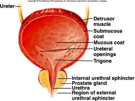
Ureters, Urinary Bladder, and Urethra
These structures transport, store, and expel urine from the body.
Ureters: Carry urine from the renal pelvis to the bladder. Peristalsis propels urine through the system.
Urinary Bladder: Stores urine; lined with transitional epithelium and contains the detrusor muscle for contraction.
Urethra: Conducts urine to the exterior; contains internal and external sphincters. There are anatomical differences between males and females.
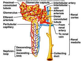
Nephron Structure and Function
Nephrons are the functional units of the kidney, responsible for filtering blood and forming urine. Each kidney contains over a million nephrons, most of which are located in the cortex (cortical nephrons), while some are juxtamedullary nephrons near the cortex-medulla junction.
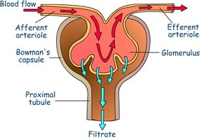
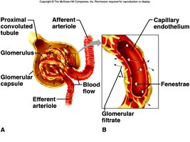
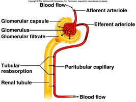
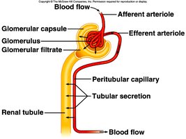
Glomerulus: Ball of capillaries specialized for filtration; blood enters via afferent arteriole and leaves via efferent arteriole.
Renal Tubule: Duct for urine collection; includes Bowman’s capsule, proximal convoluted tubule (PCT), loop of Henle, distal convoluted tubule (DCT), and collecting duct.
Peritubular Capillaries: Network of capillaries surrounding the renal tubule, involved in reabsorption and secretion.
Urine Formation: Filtration, Reabsorption, and Secretion
Urine formation occurs in the nephrons and involves three main processes:
Filtration: Occurs in the glomerulus; almost everything except proteins and cells is filtered out of the blood into Bowman’s capsule, forming filtrate.
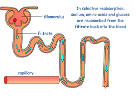
Reabsorption: Occurs primarily in the PCT; substances needed by the body (water, glucose, amino acids, electrolytes) are reabsorbed into the bloodstream via peritubular capillaries.
Secretion: The reverse of reabsorption; substances are moved from the blood into the renal tubule to be excreted as urine, primarily in the PCT (except for K+).
Summary of Filtration, Reabsorption, and Secretion
The following table summarizes the substances filtered and reabsorbed by the kidney per 24 hours:
Substance | Amount filtered (grams) | Amount reabsorbed (grams) | Amount in urine (grams) |
|---|---|---|---|
Water | 180 L | 179 L | 1 L |
Proteins | 10-20 | 10-20 | 0 |
Chlorine | 630 | 625 | 5 |
Sodium | 540 | 537 | 3 |
Bicarbonate | 180 | 180 | 0 |
Glucose | 180 | 180 | 0 |
Urea | 53 | 28 | 25 |
Potassium | 28 | 7.7 | 20.3 |
Uric acid | 8.5 | 7.7 | 0.8 |
Creatinine | 1.4 | 0 | 1.4 |
Regulation of Water, Electrolyte, and Acid-Base Balance
The kidneys play a crucial role in maintaining water, electrolyte, and acid-base balance in the blood.
Water Balance: Antidiuretic hormone (ADH) stimulates the collecting tubules to reabsorb more water.
Electrolyte Balance: Aldosterone regulates Na+ concentration; 80% of Na+ is reabsorbed in the PCT, and additional reabsorption occurs in the DCT when aldosterone is present. K+ is secreted into the filtrate.
Acid-Base Balance: Blood pH (7.35-7.45) is regulated by the lungs and kidneys. The kidneys reabsorb or secrete H+ to maintain pH.
Route of Urine Flow
After formation, urine travels through the following pathway:
Collecting duct → minor calyx → major calyx → renal pelvis → ureter → bladder → urethra → external urethral orifice
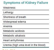
Symptoms of Kidney Failure
Kidney failure can result in a variety of symptoms due to the loss of normal kidney function.
Weakness
Lethargy
Shortness of breath
Widespread edema
Anemia
Metabolic acidosis
Metabolic alkalosis
Heart arrhythmias
Uremia (high urea level in the blood)
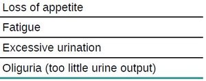
Loss of appetite
Fatigue
Excessive urination
Oliguria (too little urine output)
Color Coding Activity: Nephron Processes
Diagrams of the nephron can be used to visually represent the location and direction of key processes:
Black arrows: Filtrate formation
Red arrows: Amino acid and glucose reabsorption
Green arrows: Water movement (ADH action)
Yellow arrows: Na+ movement (aldosterone action)
Blue arrows: Tubular secretion
Additional info:
Blood flow through the nephron is essential for filtration, reabsorption, and secretion. The afferent arteriole brings blood to the glomerulus, and the efferent arteriole carries it away.
Filtrate is processed in the renal tubule, and substances are selectively reabsorbed or secreted to maintain homeostasis.
Urine composition reflects the balance of filtration, reabsorption, and secretion, and abnormal findings (e.g., glucose, blood proteins, blood cells) may indicate pathology.
