 Back
BackThe Urinary System: Structure, Function, and Regulation
Study Guide - Smart Notes
 Tailored notes based on your materials, expanded with key definitions, examples, and context.
The Urinary System
Overview and Functions
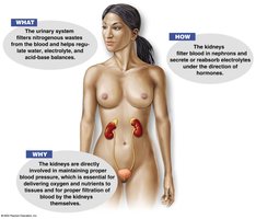
The urinary system is essential for maintaining homeostasis by regulating blood volume, blood pressure, electrolyte balance, acid-base balance, and eliminating waste products. It also plays a role in red blood cell production and vitamin D activation.
Regulation of Homeostasis: Controls blood volume, blood pressure, electrolytes, and acid-base balance.
Waste Elimination: Removes nitrogenous wastes, toxins, and drugs from the body.
Hormonal Functions: Secretes erythropoietin (EPO) for red blood cell production and activates vitamin D.
Organs of the Urinary System
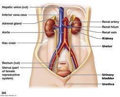
The urinary system consists of four main organs: kidneys, ureters, urinary bladder, and urethra. Each organ has a specific role in urine formation, storage, and elimination.
Kidneys: Filter blood and produce urine.
Ureters: Transport urine from kidneys to bladder.
Urinary Bladder: Stores urine until elimination.
Urethra: Conducts urine out of the body.
Kidney Anatomy and Physiology
Location and Structure
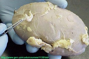
The kidneys are retroperitoneal organs located against the dorsal body wall, spanning from T12 to L3 vertebrae. They are protected by three layers: fibrous capsule, perirenal fat capsule, and renal fascia.
Renal Hilum: Indentation where ureters, blood vessels, and nerves enter/exit.
Adrenal Glands: Located atop each kidney.
Coverings: Fibrous capsule (innermost), perirenal fat capsule (middle), renal fascia (outermost).
Internal Regions of the Kidney
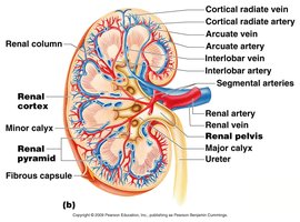
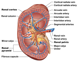
The kidney is divided into distinct regions: cortex, medulla, pyramids, columns, pelvis, and calyces. These regions facilitate urine formation and collection.
Renal Cortex: Outermost region.
Renal Medulla: Deep to cortex, contains pyramids.
Renal Pyramids: Triangular tissue regions in medulla.
Renal Columns: Separate pyramids.
Renal Pelvis: Funnel-shaped collecting tube.
Calyces: Cup-shaped structures funneling urine to pelvis.
Blood Supply and Flow
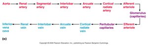
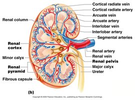
Kidneys receive a significant portion of cardiac output, with blood entering via the renal artery and exiting through the renal vein. The pathway of blood flow is highly organized to facilitate filtration.
Renal Artery: Supplies arterial blood.
Blood Flow Pathway: Aorta → Renal artery → Segmental artery → Interlobar artery → Arcuate artery → Cortical radiate artery → Afferent arteriole → Glomerulus → Efferent arteriole → Peritubular capillaries → Cortical radiate vein → Arcuate vein → Interlobar vein → Renal vein → Inferior vena cava.
Nephron Structure and Function
Nephron Anatomy
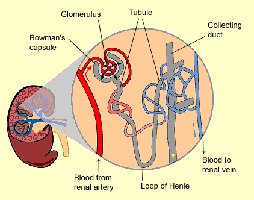
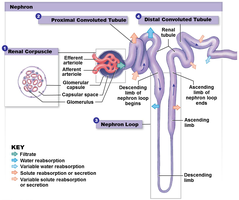
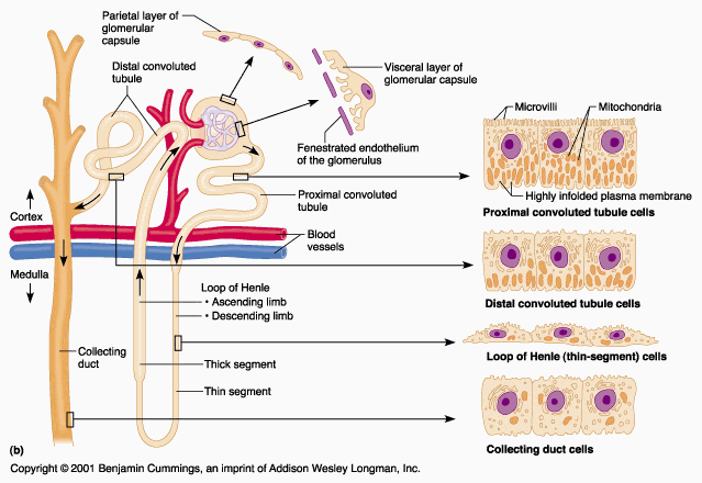
Nephrons are the functional units of the kidney, responsible for urine formation. Each nephron consists of a renal corpuscle and renal tubules.
Renal Corpuscle: Includes glomerulus and Bowman’s capsule.
Renal Tubules: Proximal convoluted tubule (PCT), nephron loop (loop of Henle), distal convoluted tubule (DCT).
Renal Corpuscle
The renal corpuscle is the site of blood filtration. The glomerulus is a capillary network surrounded by Bowman’s capsule, which collects filtrate.
Glomerulus: Specialized capillary bed for filtration.
Bowman’s Capsule: Cup-shaped structure collecting filtrate.
Renal Tubules and Collecting System
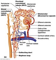
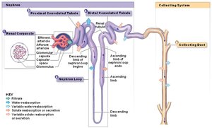
Renal tubules process filtrate through reabsorption and secretion. The collecting duct is not part of the nephron but carries urine toward the renal pelvis.
PCT: Most reabsorption occurs here.
Nephron Loop: Reabsorbs water and ions.
DCT: Variable reabsorption, influenced by hormones.
Collecting Duct: Final adjustments to urine composition.
Renal Physiology and Urine Formation
Urine Formation Steps
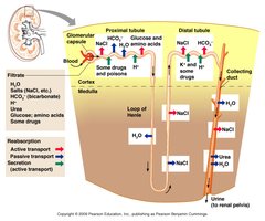
Urine is formed through three main processes: glomerular filtration, tubular reabsorption, and tubular secretion.
Glomerular Filtration: Passive process; water and solutes are filtered from blood.
Tubular Reabsorption: Useful substances are reclaimed from filtrate.
Tubular Secretion: Additional wastes and excess ions are secreted into tubules.
Glomerular Filtration
Filtration occurs in the renal corpuscle, producing filtrate similar to blood plasma but without proteins. Glomerular filtration rate (GFR) is a key measure of nephron activity.
Filtration Rate: Approximately 180 L/day.
Pressure Dependency: Filtration stops if blood pressure is too low.
Tubular Reabsorption
Most filtrate is reabsorbed, especially in the PCT. Water, glucose, amino acids, and ions are reclaimed, while nitrogenous wastes are poorly reabsorbed.
PCT: 60–70% water, nearly all organic substrates, and most sodium/chloride ions reabsorbed.
Nephron Loop: 25% water, 20–25% sodium/chloride ions reabsorbed.
DCT and Collecting Duct: Variable reabsorption, regulated by ADH and aldosterone.
Tubular Secretion
Secretion removes additional wastes and excess ions from blood into the tubules, helping maintain acid-base balance and eliminate drugs.
Materials Secreted: Hydrogen ions, potassium ions, creatinine, drugs.
Importance: Maintains acid-base balance and removes substances not filtered initially.
Fluid, Electrolyte, and Acid-Base Balance
Fluid and Electrolyte Balance
Blood composition is regulated by diet, cellular metabolism, and urine output. The kidneys play a central role in maintaining water and electrolyte balance.
Water Balance: Intake must equal output; regulated by thirst mechanism and ADH.
Electrolyte Balance: Sodium, potassium, chloride, and calcium ions are tightly regulated.
Hormonal Regulation: ADH prevents water loss; aldosterone regulates sodium and potassium.
Acid-Base Balance
Blood pH is maintained between 7.35 and 7.45. The body uses buffer systems, respiratory control, and renal mechanisms to regulate pH.
Buffer Systems: Bicarbonate, phosphate, and protein buffers.
Respiratory Control: Adjusts CO2 levels to influence pH.
Renal Mechanisms: Excretes or reabsorbs bicarbonate and hydrogen ions.
Developmental and Clinical Aspects
Developmental Aspects
Functional kidneys develop by the third month of gestation. Newborns have limited urine concentration ability, and urinary control develops with age.
Newborns: Small bladder, frequent voiding, limited concentration.
Children: Voluntary control develops by ~18 months; nighttime control by age 4.
Aging: Decreased filtration rate, increased urgency, frequency, and incontinence.
Clinical Issues
Common urinary system problems include urinary tract infections (UTIs), renal calculi, and urinary retention. Renal failure may require dialysis.
UTIs: Most common in children and elderly; E. coli is a frequent cause.
Renal Calculi: Kidney stones can obstruct urine flow.
Urinary Retention: Common in males due to prostate enlargement.
Summary Table: Normal Laboratory Values for Solutes in Plasma and Urine
Solute | Plasma (mg/dL) | Urine (mg/dL) |
|---|---|---|
Sodium (Na+) | 135–145 | 40–220 |
Potassium (K+) | 3.5–5.0 | 25–150 |
Chloride (Cl−) | 98–106 | 110–250 |
Bicarbonate (HCO3−) | 20–28 | 1.9 |
Glucose | 70–110 | 0.000 |
Urea | 8–23 | 1800 |
Creatinine | 0.6–1.5 | 140 |
Uric acid | 2–6 | 40 |
Key Equations
Carbonic Acid–Bicarbonate Buffer System:
Glomerular Filtration Rate (GFR): GFR is calculated based on creatinine clearance.
Where = urine creatinine concentration, = urine flow rate, = plasma creatinine concentration.
Conclusion
The urinary system is vital for maintaining internal balance, filtering blood, and excreting wastes. Its complex structure and regulation ensure proper function, with clinical and developmental aspects influencing health throughout life.
