 Back
BackThe Urinary System: Structure, Function, and Regulation
Study Guide - Smart Notes
 Tailored notes based on your materials, expanded with key definitions, examples, and context.
The Urinary System
Overview and Functions
The urinary system is essential for maintaining homeostasis by filtering blood, removing waste, and regulating fluid and electrolyte balance. It consists of the kidneys, ureters, urinary bladder, and urethra.
Excretion: Removal of metabolic waste products, especially urea.
Elimination: Discharge of waste products from the body.
Homeostatic Regulation: Maintains blood pH, ion concentrations, and fluid volume.
Other Functions: Conservation of nutrients, regulation of erythrocyte production, urine storage, and excretion.
Kidney Anatomy and Location
Gross Anatomy
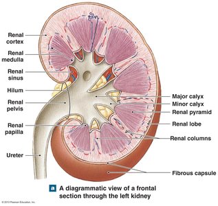
The kidneys are retroperitoneal organs located on the posterior abdominal wall, adjacent to the lower vertebrae. Each kidney is covered by a fibrous capsule and surrounded by adipose tissue for protection.
Cortex: The outer, lighter region of the kidney.
Medulla: The inner, darker region containing renal pyramids.
Renal Pyramids: Triangular structures in the medulla.
Renal Papilla: The apex of each pyramid, where urine drains into minor calyces.
Renal Columns: Extensions of cortical tissue between pyramids.
Hilum: The entry/exit site for vessels, nerves, and ureter.
Renal Sinus: Cavity within the kidney that houses the renal pelvis, calyces, and vessels.
Minor and Major Calyces: Collect urine from pyramids and funnel it to the renal pelvis.
Renal Pelvis: Central collecting region for urine before it enters the ureter.
Blood Flow in the Kidneys
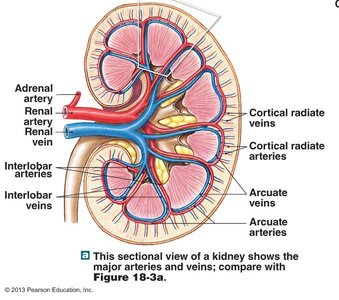
The kidneys receive 20–25% of cardiac output. Blood flows through a series of arteries and veins, closely associated with nephron function.
Renal artery → Interlobar artery → Arcuate artery → Cortical radiate artery → Afferent arteriole → Glomerular capillaries → Efferent arteriole → Peritubular capillaries → Cortical radiate vein → Arcuate vein → Interlobar vein → Renal vein
Nephron Structure and Types
Nephron Anatomy
The nephron is the basic functional unit of the kidney, responsible for urine formation. Each kidney contains approximately one million nephrons.
Renal Corpuscle: Consists of the glomerulus (a cluster of capillaries) and the glomerular capsule (Bowman's capsule) that surrounds it.
Renal Tubule: Includes the proximal convoluted tubule, loop of Henle, distal convoluted tubule, and collecting duct.
Types of Nephrons
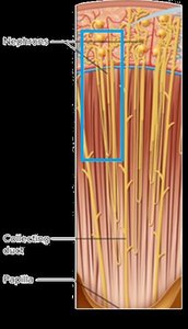
Cortical Nephrons: Located near the outer cortex; have short loops of Henle.
Juxtamedullary Nephrons: Located near the cortex-medulla border; have long loops of Henle, important for concentrating urine.
Pathway of Urine Formation and Excretion
Pathway
Urine is formed through a series of steps as blood is filtered and processed by the nephron:
Glomerular capsule
Proximal convoluted tubule
Loop of Henle
Distal convoluted tubule
Collecting duct
Papillary duct
Renal papilla
Minor calyx
Major calyx
Renal pelvis
Ureter
Bladder
Urethra
Out of body
Urine Formation: The Three Main Processes
1. Glomerular Filtration
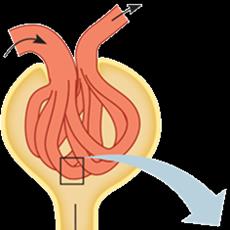
Filtration occurs in the renal corpuscle, where blood pressure forces water and solutes from the glomerulus into the glomerular capsule, forming filtrate.
Glomerular Filtration Rate (GFR): The rate at which filtrate is produced; normal GFR is 61–104 mL/min/1.73 m2.
Filtration Pressures: Determined by glomerular hydrostatic pressure, colloid osmotic pressure, and capsular hydrostatic pressure.
Net Filtration Pressure (NFP):
2. Tubular Reabsorption
Reabsorption is the process by which water and essential solutes are reclaimed from the filtrate and returned to the blood, primarily in the proximal convoluted tubule.
3. Tubular Secretion
Secretion involves the active transport of additional wastes and excess ions from the blood into the renal tubule for excretion.
Regulation of Glomerular Filtration Rate (GFR)
Mechanisms of Regulation
Renal Autoregulation: The kidney self-adjusts blood flow and filtration rate.
Neural Regulation: The nervous system can constrict afferent arterioles, reducing GFR during stress or blood loss.
Hormonal Regulation: The renin-angiotensin-aldosterone system (RAAS) adjusts GFR in response to blood pressure and volume changes.
Consequences of Abnormal GFR
Hyperfiltration (High GFR)
GFR > 130 mL/min/1.73 m2
Substances are not adequately reabsorbed, leading to excessive loss of water and solutes.
Associated with diseases such as diabetes mellitus and hypertension.
Hypofiltration (Low GFR)
GFR < 60 mL/min/1.73 m2
Nearly all substances are reabsorbed, and waste products may not be adequately excreted.
Associated with chronic kidney disease and other renal pathologies.
Summary Table: Key Structures and Functions
Structure | Function |
|---|---|
Kidney | Filters blood, forms urine |
Nephron | Functional unit; filtration, reabsorption, secretion |
Glomerulus | Filtration of blood plasma |
Proximal Convoluted Tubule | Reabsorption of water, ions, nutrients |
Loop of Henle | Concentration of urine |
Distal Convoluted Tubule | Secretion and selective reabsorption |
Collecting Duct | Final concentration of urine |
Key Terms and Concepts
Excretion: Removal of metabolic wastes from the body.
Filtration: Movement of water and solutes from blood into the nephron.
Reabsorption: Return of water and solutes from filtrate to blood.
Secretion: Addition of substances from blood into the filtrate.
GFR: Glomerular Filtration Rate, a measure of kidney function.
Renin-Angiotensin-Aldosterone System (RAAS): Hormonal system regulating blood pressure and GFR.
Additional info: The maintenance of GFR is critical for homeostasis. Both hyperfiltration and hypofiltration can lead to significant health issues, including fluid and electrolyte imbalances, accumulation of toxins, and progression of kidney disease.
