 Back
BackThe Urinary System: Structure, Function, and Physiology
Study Guide - Smart Notes
 Tailored notes based on your materials, expanded with key definitions, examples, and context.
The Urinary System
Overview and Functions
The urinary system is essential for maintaining the body's internal environment by regulating water, solute concentrations, and removing metabolic wastes. It consists of the kidneys, ureters, urinary bladder, and urethra.
Regulation of water and solutes: Kidneys control total water volume and solute concentration.
Ion balance: Maintains ion concentrations in extracellular fluid.
Acid-base balance: Ensures long-term acid-base equilibrium.
Excretion: Removes metabolic wastes, toxins, and drugs.
Hormone production: Produces erythropoietin (stimulates red blood cell production) and renin (regulates blood pressure).
Vitamin D activation: Converts vitamin D to its active form.
Gluconeogenesis: Generates glucose during prolonged fasting.
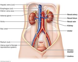
Components of the Urinary System
Kidneys: Major excretory organs.
Ureters: Transport urine from kidneys to bladder.
Urinary bladder: Temporary storage for urine.
Urethra: Conducts urine out of the body.
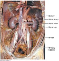
Gross Anatomy of the Kidneys
Location and External Anatomy
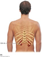
The kidneys are retroperitoneal, located in the superior lumbar region between T12 and L5. The right kidney is slightly lower due to the liver. Each kidney is topped by an adrenal gland.
Renal hilum: Entry/exit for ureters, blood vessels, lymphatics, and nerves.
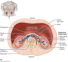
Supportive tissue layers:
Renal fascia: Anchoring outer layer.
Perirenal fat capsule: Cushions the kidney.
Fibrous capsule: Prevents infection spread.
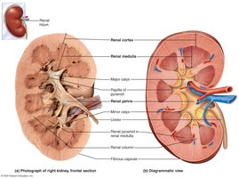
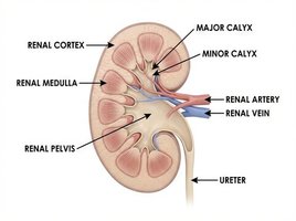
Internal Gross Anatomy
The kidney has three main regions:
Renal cortex: Superficial, granular region.
Renal medulla: Deep region with cone-shaped pyramids.
Renal pelvis: Funnel-shaped tube continuous with ureter.
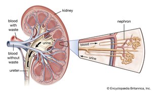
Urine flow: Renal pyramid → minor calyx → major calyx → renal pelvis → ureter.
Blood and Nerve Supply
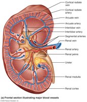
Renal Circulation
Kidneys receive about one-fourth of cardiac output per minute.
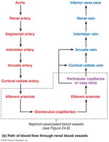
Arterial flow: Renal → segmental → interlobar → arcuate → cortical radiate (interlobular).
Venous flow: Cortical radiate → arcuate → interlobar → renal veins.
Nerve supply: Sympathetic fibers from renal plexus.
Nephrons: The Functional Units
Structure of Nephrons
Nephrons are the structural and functional units that form urine. Each kidney contains over one million nephrons.
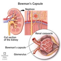
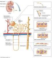
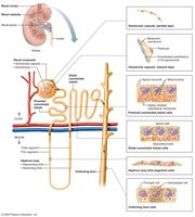
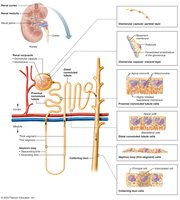
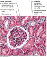
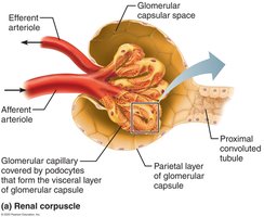
Renal corpuscle: Includes the glomerulus and glomerular (Bowman's) capsule.
Renal tubule: Includes proximal convoluted tubule, nephron loop, distal convoluted tubule, and collecting duct.
Renal Corpuscle
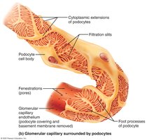
Glomerulus: Tuft of fenestrated capillaries for efficient filtrate formation.
Bowman's capsule: Cup-shaped structure surrounding the glomerulus.
Renal Tubule and Collecting Duct
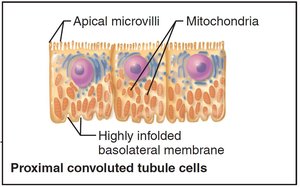
Proximal convoluted tubule (PCT): Cuboidal cells with dense microvilli for reabsorption and secretion.
Nephron loop (Loop of Henle): U-shaped, with descending and ascending limbs.
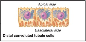
Distal convoluted tubule (DCT): Cuboidal cells, mainly for secretion.
Collecting duct: Receives filtrate from DCT.
Classes of Nephrons
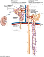
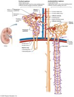
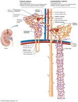
Cortical nephrons: 85% of nephrons, mostly in cortex.
Juxtamedullary nephrons: Long loops deeply invade medulla, crucial for concentrated urine production.
Nephron Capillary Beds
Types of Capillary Beds
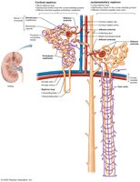
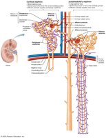
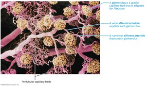
Glomerulus: Specialized for filtration, fed and drained by arterioles.
Peritubular capillaries: Low-pressure, porous, adapted for absorption.
Vasa recta: Long vessels parallel to nephron loops in juxtamedullary nephrons, important for urine concentration.
Juxtaglomerular Complex (JGC)
Structure and Function
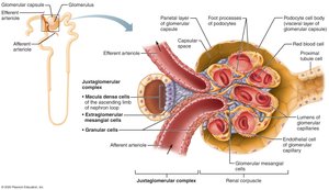
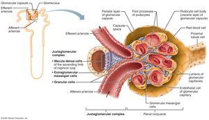
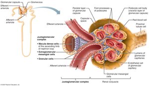
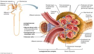
The JGC regulates filtrate formation and blood pressure. It consists of:
Macula densa: Chemoreceptors sensing NaCl content.
Granular cells: Mechanoreceptors sensing blood pressure, secrete renin.
Extraglomerular mesangial cells: Pass signals between macula densa and granular cells.
Physiology of the Kidney
Urine Formation
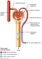
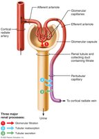
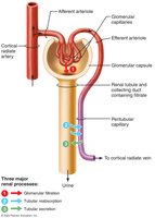
The kidneys process 180L of fluid daily, forming only 1.5L of urine. Three main processes are involved:
Glomerular filtration: Produces cell- and protein-free filtrate.
Tubular reabsorption: Returns 99% of substances to blood.
Tubular secretion: Moves substances from blood to filtrate.
Glomerular Filtration
Passive process: Driven by hydrostatic pressure.
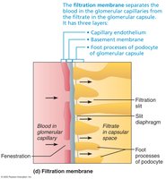
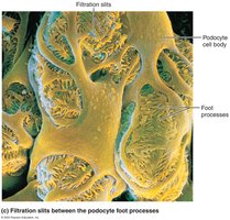
Filtration membrane: Three layers: fenestrated endothelium, basement membrane, and podocyte foot processes.
Allows passage: Water, glucose, amino acids, nitrogenous wastes; blocks cells and most proteins.
Pressures Affecting Filtration
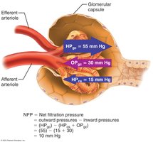
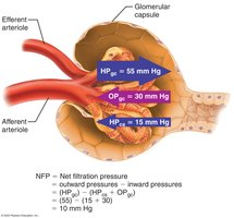
Outward pressure: Hydrostatic pressure in glomerular capillaries (HPgc), ~55 mmHg.
Inward pressures: Hydrostatic pressure in capsular space (HPcs), 15 mmHg; colloid osmotic pressure in capillaries (OPgc), 30 mmHg.
Net filtration pressure (NFP):
Glomerular Filtration Rate (GFR)
Definition: Volume of filtrate formed per minute by both kidneys (120–125 ml/min).
Factors: NFP, surface area, membrane permeability.
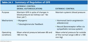
Regulation of Glomerular Filtration
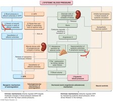
Intrinsic controls: Renal autoregulation (myogenic and tubuloglomerular feedback mechanisms).
Extrinsic controls: Neural and hormonal mechanisms (renin-angiotensin-aldosterone system).
Tubular Reabsorption and Secretion
Tubular Reabsorption
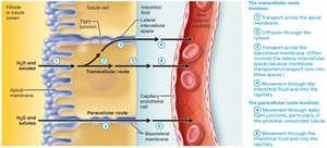
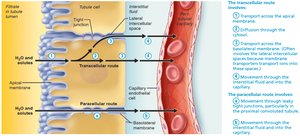
Tubular reabsorption reclaims most of the filtrate and returns it to the blood.
Routes: Transcellular (through cells) and paracellular (between cells).
Active and passive transport: Used for water and solutes.
Transport Maximum
Definition: Maximum rate of reabsorption for each substance, determined by number of carriers.
Clinical relevance: Hyperglycemia can exceed Tm, causing glucose in urine.
Reabsorptive Capabilities of Renal Tubules
PCT: Site of most reabsorption (all nutrients, 65% Na+ and water).
Nephron loop: Descending limb allows water reabsorption; ascending limb allows solute reabsorption.
DCT and collecting duct: Reabsorption regulated by hormones (ADH, aldosterone, atrial natriuretic peptide, parathyroid hormone).
Tubular Secretion
Tubular secretion moves substances from blood into filtrate, mainly in the PCT.
Functions: Disposal of drugs, elimination of reabsorbed wastes, removal of excess K+, regulation of blood pH.
Regulation of Urine Concentration and Volume
Countercurrent Mechanisms
The kidneys use countercurrent mechanisms to maintain a medullary osmotic gradient, allowing concentration or dilution of urine.
Countercurrent multiplier: Interaction of filtrate flow in nephron loops.
Countercurrent exchanger: Blood flow in vasa recta preserves gradient.
Formation of Dilute or Concentrated Urine
Overhydration: Produces dilute urine (low ADH).
Dehydration: Produces concentrated urine (high ADH).
Urea Recycling
Urea contributes to the medullary osmotic gradient by recycling between nephron loop and collecting duct.
Clinical Evaluation of Kidney Function
Urinalysis and Renal Clearance
Urinalysis: Examines urine for disease or illegal substances.
Renal clearance: Volume of plasma cleared of a substance per unit time. Used to assess GFR and detect renal disease.
Formula: where C = clearance rate, U = urine concentration, V = urine flow rate, P = plasma concentration.
Clinical Disorders
Chronic renal disease: GFR < 60 ml/min for 3 months.
Renal failure: GFR < 15 ml/min; causes uremia, treated by dialysis or transplant.
Urine: Composition and Characteristics
Chemical Composition
95% water, 5% solutes (urea, uric acid, creatinine, ions).
Abnormal components may indicate pathology.
Physical Characteristics
Color: Pale to deep yellow; abnormal colors may indicate disease.
Odor: Slightly aromatic; changes with disease or diet.
pH: Slightly acidic (~6), varies with diet.
Specific gravity: 1.001–1.035.
Transport, Storage, and Elimination of Urine
Ureters
Ureters are muscular tubes that convey urine from kidneys to bladder.
Three layers: Mucosa (transitional epithelium), muscularis (smooth muscle), adventitia (fibrous connective tissue).
Peristalsis: Propels urine; rate adjusted to urine formation.
Clinical Disorders: Renal Calculi
Kidney stones: Crystallized salts; can block ureter and cause pain.
Treatment: Lithotripsy, hydration, surgical removal.
Urinary Bladder
Muscular sac: Temporary storage for urine.
Trigone: Area outlined by ureter and urethra openings; infection-prone.
Wall layers: Mucosa, detrusor muscle, adventitia.
Capacity: Moderately full bladder holds ~500 ml; can expand further.
Urethra
Muscular tube: Drains bladder.
Lining: Varies from transitional to stratified squamous epithelium.
Sphincters: Internal (involuntary) and external (voluntary).
Female urethra: Short, tightly bound to anterior vaginal wall.
Male urethra: Longer, carries urine and semen; divided into prostatic, membranous, and spongy regions.
Clinical Disorders: Urinary Tract Infections (UTIs)
Causes: Improper hygiene, short female urethra.
Symptoms: Pain, frequent urination, blood, cramping.
Treatment: Antibiotics.
Other Urinary Problems
Urethritis: Inflammation of urethra.
Cystitis: Inflammation of bladder.
Pyelitis/Pyelonephritis: Inflammation of kidneys.
Symptoms: Dysuria, urgency, fever, cloudy/bloody urine, back pain.
Micturition (Urination)
Mechanism
Micturition involves contraction of detrusor muscle, opening of internal and external sphincters.
Storage phase: Bladder fills with urine.
Voiding phase: Bladder contracts, sphincters open, urine expelled.
Reflexive and Voluntary Control
Infants: Reflexive urination via spinal stretch receptors.
Adults: Pontine centers mature, allowing voluntary control.
Clinical Problems
Incontinence: Loss of bladder control; stress or overflow types.
Retention: Inability to expel urine; may require catheterization.
Additional info: Academic context and explanations have been expanded for clarity and completeness. All included images are directly relevant to the adjacent content, reinforcing anatomical and physiological concepts.
