 Back
CH 4: Tissue Level of Organization: Epithelial and Connective Tissues
Study Guide - Smart Notes
 Tailored notes based on your materials, expanded with key definitions, examples, and context.
Ch 4: The Four Major Types of Tissues
Overview of Tissue Types
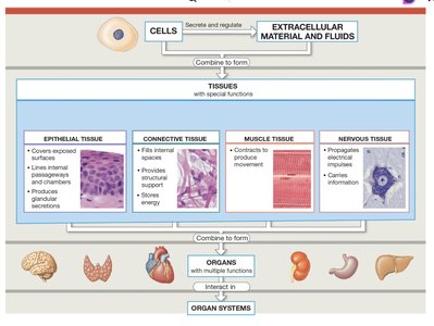
The human body is composed of trillions of cells organized into four primary tissue types, each with specialized functions. The study of tissues is known as histology. These tissues combine to form organs and organ systems, enabling complex physiological functions.
Epithelial Tissue: Covers exposed surfaces, lines internal passageways and chambers, and forms glands.
Connective Tissue: Fills internal spaces, provides structural support, transports materials, and stores energy.
Muscle Tissue: Specialized for contraction, enabling movement.
Nervous Tissue: Carries information via electrical impulses.
Epithelial Tissue
Functions and Characteristics
Epithelial tissue forms the body's external and internal surfaces and is essential for protection, permeability, sensation, and secretion. It consists of tightly packed cells with distinct structural features.
Physical Protection: Shields surfaces from abrasion, dehydration, and chemical/biological agents.
Control Permeability: Regulates entry and exit of substances; can be modified by hormones or physical stress.
Provide Sensation: Rich in sensory nerves; includes specialized neuroepithelia for senses like smell and taste.
Produce Specialized Secretions: Gland cells release substances for protection, regulation, or communication.
Key Structural Features
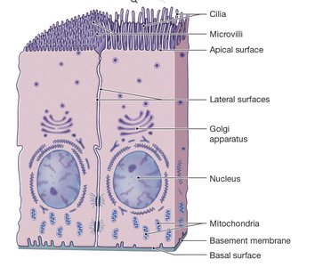
Polarity: Distinct apical (exposed) and basal (attached) surfaces.
Cellularity: Cells are closely bound by cell junctions.
Attachment: Bound to a basement membrane composed of basal and reticular lamina.
Avascularity: Lacks blood vessels; nutrients diffuse from adjacent tissues.
Regeneration: High rate of cell division and replacement via stem cells.
Specializations of Epithelial Cells
Microvilli: Increase surface area for absorption and secretion, especially in digestive and kidney epithelia.
Cilia: Motile structures that move substances across surfaces, such as mucus in the respiratory tract.
Maintaining Epithelial Integrity
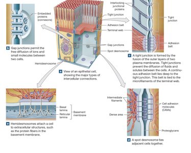
Intercellular Connections: Cell junctions (gap, tight, desmosomes) and cell adhesion molecules (CAMs) ensure cohesion and communication.
Attachment to Basement Membrane: Provides structural support and selective filtration.
Maintenance and Repair: Continuous stem cell division replaces damaged cells.
Classification of Epithelia
Cell Shape and Layering
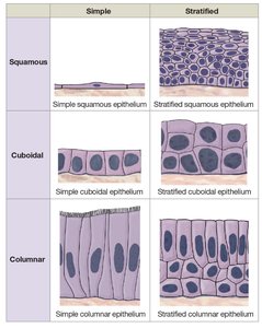
Epithelia are classified by cell shape and the number of layers:
Cell Shapes:
Squamous: Thin, flat cells.
Cuboidal: Box-like cells.
Columnar: Tall, slender cells.
Layering:
Simple: Single layer; found in protected areas and where absorption/secretion occurs.
Stratified: Multiple layers; found in areas exposed to stress.
Squamous Epithelia
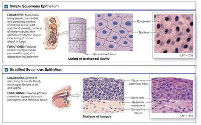
Simple Squamous: Delicate, lines protected regions (alveoli, body cavities, heart, blood vessels). Specialized as mesothelium (body cavities) and endothelium (heart, vessels).
Stratified Squamous: Multiple layers, protects against mechanical stress; keratinized (skin) or nonkeratinized (mouth, esophagus).
Cuboidal and Transitional Epithelia
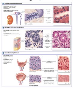
Simple Cuboidal: Limited protection, secretion/absorption; found in glands and kidney tubules.
Stratified Cuboidal: Rare, found in sweat gland ducts and mammary glands.
Transitional Epithelium: Stratified, tolerates stretching; found in urinary bladder.
Columnar Epithelia
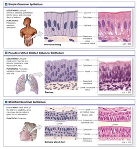
Simple Columnar: Absorption/secretion; found in intestines, stomach.
Pseudostratified Columnar: Appears layered, all cells touch basement membrane; often ciliated, found in respiratory tract.
Stratified Columnar: Rare, provides protection in pharynx, urethra, and large ducts.
Glandular Epithelia
Types of Glands
Endocrine Glands: Ductless, release hormones into blood (e.g., thyroid, pituitary).
Exocrine Glands: Release secretions onto surfaces or into ducts (e.g., sweat, salivary glands).
Exocrine Gland Structure
Unicellular: Single goblet cells secrete mucin.
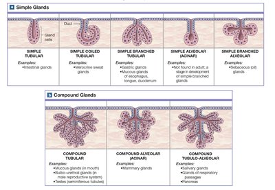
Multicellular: Secretory sheets or glands with ducts; classified as simple (single duct) or compound (branched ducts).
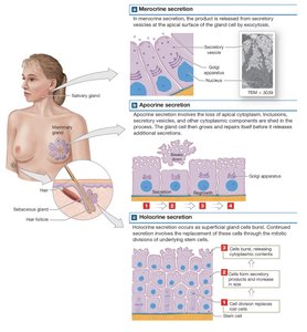
Methods of Secretion
Merocrine: Exocytosis; cell remains intact (e.g., sweat glands).
Apocrine: Loss of apical cytoplasm; cell repairs itself (e.g., mammary glands).
Holocrine: Cell bursts, releasing contents; cell is destroyed (e.g., sebaceous glands).
Types of Exocrine Secretions
Serous: Watery, enzyme-rich (e.g., parotid glands).
Mucous: Mucin-rich, forms mucus (e.g., sublingual glands).
Mixed: Both serous and mucous (e.g., submandibular glands).
Connective Tissue
Functions and Categories
Connective tissue supports, connects, and protects other tissues. It consists of specialized cells, extracellular fibers, and ground substance, forming the matrix. Connective tissues are highly vascular and contain sensory receptors.
Establishes structural framework
Transports fluids and dissolved materials
Protects delicate organs
Supports and interconnects tissues
Stores energy (triglycerides)
Defends against microorganisms
Three main categories:
Connective Tissue Proper: Loose (e.g., adipose) and dense (e.g., tendons).
Fluid Connective Tissues: Blood and lymph.
Supporting Connective Tissues: Cartilage and bone.
Fluid Connective Tissues: Blood and Lymph
Blood
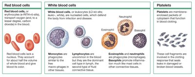
Blood is a fluid connective tissue with a matrix called plasma. It contains three types of formed elements:
Red Blood Cells (Erythrocytes): Transport oxygen and carbon dioxide; lack nuclei.
White Blood Cells (Leukocytes): Defend against infection and disease; include monocytes, lymphocytes, eosinophils, neutrophils, and basophils.
Platelets: Cell fragments involved in blood clotting.
Lymph
Lymph forms as interstitial fluid enters lymphatic vessels. It is monitored by immune cells and returned to the bloodstream, maintaining homeostasis by regulating fluid levels and alerting the immune system to infection.
Recirculation: Fluid moves from blood (plasma) to interstitial fluid to lymph and back to blood.
Homeostasis: Maintains nutrient, waste, and toxin balance; supports immune function.
Additional info: Connective tissue proper, fluid, and supporting connective tissues are discussed in greater detail in subsequent chapters.
