 Back
BackTissue: The Living Fabric – Structure and Function of Human Tissues
Study Guide - Smart Notes
 Tailored notes based on your materials, expanded with key definitions, examples, and context.
Tissue: The Living Fabric
Introduction to Tissues
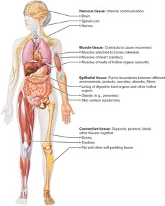
Tissues are groups of cells similar in structure that perform common or related functions, maintaining homeostasis in the body. The study of tissues is called histology. There are four basic tissue types in the human body: epithelial, connective, muscle, and nervous tissue.
Epithelial Tissue
Definition and Main Categories
Epithelial tissue (epithelium) is a sheet of cells that covers body surfaces or lines body cavities. It exists in two main forms:
Covering and lining epithelium: Forms the outer layer of the skin and lines open cavities and organs.
Glandular epithelium: Forms glands, such as salivary glands.
Main functions include protection, absorption, filtration, excretion, secretion, and sensory reception.
Special Characteristics of Epithelial Tissues
Polarity: Cells have an apical (top) surface and a basal (bottom) surface, each with distinct structures and functions.
Specialized contacts: Cells are tightly joined by junctions such as tight junctions and desmosomes.
Supported by connective tissue: All epithelial sheets rest on a basement membrane composed of basal and reticular lamina.
Avascular but innervated: Epithelia lack blood vessels but are supplied by nerve fibers; nutrients diffuse from underlying tissues.
Regeneration: High regenerative capacity, especially in areas exposed to friction or hostile environments.

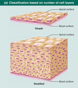
Classification of Epithelial Tissue
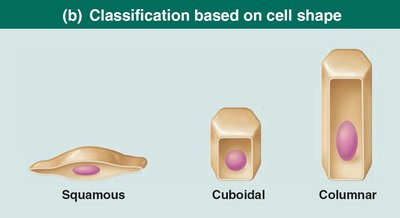
Epithelia are classified by the number of cell layers and cell shape:
Simple epithelia: Single cell layer; functions in absorption, secretion, and filtration.
Stratified epithelia: Two or more layers; found in high-abrasion areas for protection.
Cell shapes include:
Squamous: Flattened and scale-like
Cuboidal: Box-like
Columnar: Tall and column-shaped
Types of Epithelial Tissue
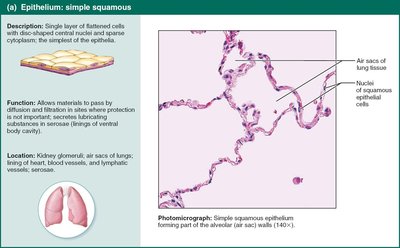
Simple Squamous Epithelium: Thin, allows diffusion/filtration; found in lungs, kidneys, blood vessels.
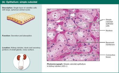
Simple Cuboidal Epithelium: Secretion and absorption; found in kidney tubules, ducts.
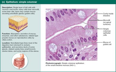
Simple Columnar Epithelium: Absorption, secretion; lines digestive tract, gallbladder.
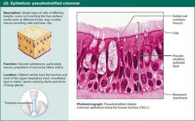
Pseudostratified Columnar Epithelium: Appears layered but is single-layered; secretion and movement of mucus; found in respiratory tract.
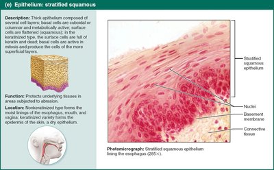
Stratified Squamous Epithelium: Protection; found in skin, mouth, esophagus.
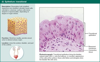
Transitional Epithelium: Stretches; lines urinary bladder, ureters.
Glandular Epithelia
A gland consists of one or more cells that secrete an aqueous fluid (secretion). Glands are classified by:
Site of product release: Endocrine (ductless, hormones) vs. Exocrine (ducts, onto surfaces)
Number of cells: Unicellular (e.g., goblet cells) vs. Multicellular (e.g., salivary glands)
Connective Tissue
Overview and Functions
Connective tissue is the most abundant and widely distributed tissue type. Its major functions include binding and support, protection, insulation, energy storage, and transportation of substances (e.g., blood).
Four main classes:
Connective tissue proper
Cartilage
Bone
Blood
Comparison of Classes of Connective Tissues
Tissue Class | Subclasses | Cells | Matrix | General Features |
|---|---|---|---|---|
Connective Tissue Proper | Loose (areolar, adipose, reticular); Dense (regular, irregular, elastic) | Fibroblasts, fibrocytes, defense cells, adipocytes | Gel-like ground substance; collagen, reticular, elastic fibers | Binding, resisting tension, water/salt reservoir, energy storage |
Cartilage | Hyaline, elastic, fibrocartilage | Chondroblasts, chondrocytes | Gel-like ground substance; collagen, elastic fibers | Resists compression, supports, avascular |
Bone | Compact, spongy | Osteoblasts, osteocytes | Calcified ground substance; collagen fibers | Hard, supports, resists tension/compression |
Blood | --- | RBCs, WBCs, platelets | Plasma (fluid); no fibers | Transport of gases, nutrients, wastes |
Common Characteristics and Elements
Extracellular matrix: Nonliving material that supports cells, allowing them to bear weight and withstand tension.
Common origin: All connective tissues arise from embryonic mesenchyme.
Three main components: ground substance, fibers (collagen, elastic, reticular), and cells (blasts and cytes).
Types of Connective Tissue Proper
Loose connective tissue: Areolar (packing material), adipose (fat storage), reticular (supports blood cells in lymphoid organs).
Dense connective tissue: Dense regular (tendons, ligaments), dense irregular (dermis), elastic (arteries, vertebral ligaments).
Cartilage, Bone, and Blood
Cartilage: Hyaline (joints, ribs), elastic (ear), fibrocartilage (intervertebral discs).
Bone: Supports, protects, stores fat, synthesizes blood cells.
Blood: Fluid tissue for transport.
Muscle Tissue
Types and Functions
Muscle tissue is highly vascularized and responsible for movement. Muscle cells contain myofilaments (actin and myosin) for contraction. Three types:
Skeletal muscle: Voluntary, striated, multinucleated; moves bones.
Cardiac muscle: Involuntary, striated, one nucleus, intercalated discs; found in heart.
Smooth muscle: Involuntary, non-striated, spindle-shaped; found in walls of hollow organs.
Nervous Tissue
Structure and Function
Nervous tissue is the main component of the nervous system (brain, spinal cord, nerves). It regulates and controls body functions. Two main cell types:
Neurons: Respond to stimuli and transmit electrical signals via dendrites and axons.
Glial cells (neuroglia): Support, insulate, and protect neurons.
Membranes
Types of Membranes
Cutaneous membrane: Skin; keratinized stratified squamous epithelium attached to connective tissue; dry membrane.
Mucous membranes: Line body cavities open to exterior (digestive, respiratory, urogenital tracts); moist, may secrete mucus.
Serous membranes: Line closed ventral body cavities; simple squamous epithelium (mesothelium) on areolar connective tissue; secrete serous fluid. Parietal serosae line cavity walls, visceral serosae cover organs.
Tissue Repair
Process of Tissue Repair
When tissues are damaged, repair occurs via:
Regeneration: Replacement by the same kind of tissue, restoring function.
Fibrosis: Replacement by connective tissue (scar), function may be lost.
Steps:
Inflammation: Release of chemicals, dilation of blood vessels, increased permeability, clotting.
Organization: Blood clot replaced by granulation tissue, epithelium regenerates, fibroblasts bridge gap.
Regeneration and fibrosis: Scab detaches, tissue matures, scar may remain.
Regenerative Capacity of Tissues
High: Epithelial, bone, areolar, dense irregular, blood-forming tissue
Moderate: Smooth muscle, dense regular connective tissue
Low/None: Cardiac muscle, nervous tissue in brain/spinal cord
Developmental Aspects of Tissues
Primary germ layers (ectoderm, mesoderm, endoderm) form early in embryonic development and give rise to all tissues:
Nerve tissue: Ectoderm
Muscle and connective tissue: Mesoderm
Epithelial tissue: All three germ layers
With aging, tissue repair is less efficient, epithelia thin, and risk of cancer increases due to DNA mutations.
