 Back
Tissues: Structure, Classification, and Function in the Human Body
Study Guide - Smart Notes
 Tailored notes based on your materials, expanded with key definitions, examples, and context.
Tissues: An Introduction
Definition and Overview
Tissues are groups of cells with similar structure that work together to perform a common function. In the human body, tissues are the fundamental units that organize cells into functional groups, allowing for complex biological processes.
Tissue: A group of cells of similar structure performing a common function.
Cells in tissues are often specialized and work together in a coordinated manner.
Four Basic Tissue Types and Their Functions
Epithelial tissue: Covers body surfaces and lines cavities; involved in protection, secretion, absorption, and filtration.
Connective tissue: Supports, binds, and protects other tissues and organs; includes bone, cartilage, blood, and connective tissue proper.
Muscle tissue: Responsible for movement; includes skeletal, cardiac, and smooth muscle.
Nervous tissue: Controls and communicates; found in the brain, spinal cord, and nerves.
Epithelial Tissue
General Characteristics
Epithelial tissue forms the covering or lining of all internal and external body surfaces and constitutes the functional tissue of glands.
Functions: Protection, secretion, absorption, diffusion, filtration, and sensory reception.
Forms parts of most glands.
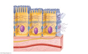
Special Characteristics of Epithelia
Cellularity: Composed almost entirely of tightly packed cells with minimal extracellular material.
Specialized contacts: Cells are joined by specialized junctions (tight junctions, desmosomes, gap junctions).
Polarity: Apical (top) surface differs from basal (bottom) surface in structure and function.
Support by connective tissue: All epithelial sheets rest upon and are supported by connective tissue.
Avascular but innervated: Contains no blood vessels but is supplied by nerve fibers; nutrients diffuse from underlying connective tissue.
Regeneration: High capacity for renewal through cell division.
Classification of Epithelia
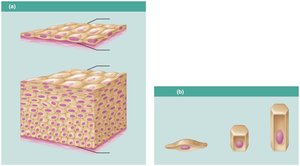
Epithelia are classified based on the number of cell layers and the shape of the cells at the apical surface.
Number of layers:
Simple: Single layer of cells attached to the basement membrane.
Stratified: Multiple layers; basal layer attached to basement membrane.
Cell shape:
Squamous: Cells are wider than tall (flat, disc-like).
Cuboidal: Cells are as wide as tall (cube-shaped).
Columnar: Cells are taller than wide (column-shaped).
Types of Epithelia: Structure, Function, and Location
Simple Epithelia
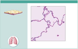
Simple squamous epithelium:
Description: Single layer of flat cells with disc-shaped nuclei.
Function: Allows passage of materials by diffusion and filtration; secretes lubricating substances.
Location: Kidney glomeruli, air sacs of lungs, lining of heart, blood vessels, lymphatic vessels, serosae.
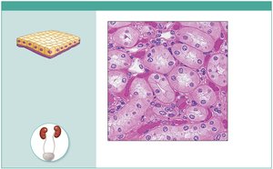
Simple cuboidal epithelium:
Description: Single layer of cube-like cells with large, spherical nuclei.
Function: Secretion and absorption.
Location: Kidney tubules, ducts and secretory portions of small glands, ovary surface.
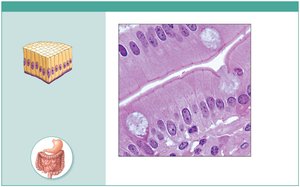
Simple columnar epithelium:
Description: Single layer of tall, column-shaped cells; may have cilia or microvilli; may contain goblet cells.
Function: Absorption; secretion of mucus, enzymes, and other substances; ciliated type propels mucus or reproductive cells.
Location: Nonciliated: digestive tract, gallbladder, ducts of some glands; ciliated: small bronchi, uterine tubes, uterus.
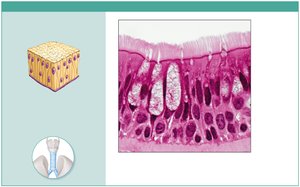
Pseudostratified columnar epithelium:
Description: Single layer of cells of varying heights; nuclei at different levels; may have cilia and goblet cells.
Function: Secretion of mucus; propulsion of mucus by cilia.
Location: Ciliated: trachea, upper respiratory tract; nonciliated: male reproductive ducts, ducts of large glands.
Stratified Epithelia
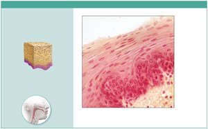
Stratified squamous epithelium:
Description: Multiple layers; basal cells cuboidal or columnar, surface cells squamous; keratinized or nonkeratinized.
Function: Protects underlying tissues in areas subject to abrasion.
Location: Keratinized: epidermis; nonkeratinized: esophagus, mouth, anus, vagina, urethra.
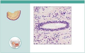
Stratified cuboidal epithelium:
Description: Generally two layers of cube-shaped cells.
Function: Protection.
Location: Ducts of mammary glands, salivary glands, largest sweat glands.
Stratified columnar epithelium:
Description: Several layers; basal cells cuboidal, superficial cells columnar.
Function: Protection and secretion.
Location: Rare; male urethra, large ducts of some glands.
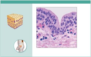
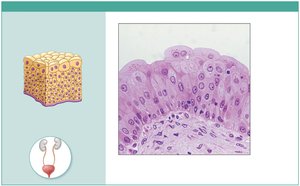
Transitional epithelium:
Description: Resembles both stratified squamous and cuboidal; surface cells dome-shaped or squamous depending on stretch.
Function: Permits distension of urinary organs.
Location: Ureters, bladder, proximal urethra.
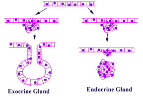
Glands: Endocrine and Exocrine
Endocrine glands: Ductless; secrete hormones directly into tissue fluid or blood. Example: Thyroid gland.
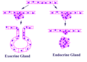
Exocrine glands: Secrete products into ducts that open onto epithelial surfaces. Examples: Sweat, oil, salivary glands, liver, pancreas.
Lateral Surface Features—Cell Junctions
Tight junctions (zona occludens): Impermeable junctions that seal cells together, preventing passage of molecules between cells.
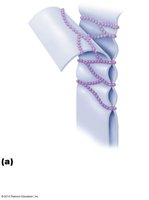
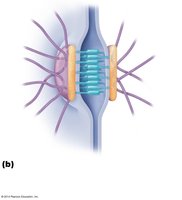
Desmosomes: Anchoring junctions that bind cells together, providing mechanical stability.
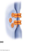
Gap junctions: Communicating junctions allowing ions and small molecules to pass between cells; important for intercellular communication.
Apical Surface Features
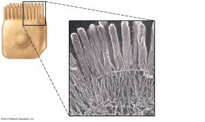
Microvilli: Fingerlike extensions of the plasma membrane that increase surface area for absorption; abundant in kidney tubules and small intestine.
Cilia: Whiplike, motile extensions that move substances across the cell surface; found in respiratory tract and uterine tubes.
Connective Tissue
Overview and Functions
Connective tissue is the most diverse and abundant tissue type, providing support, binding, and protection for organs and tissues throughout the body.
Main classes: Connective tissue proper, cartilage, bone tissue, blood.
Functions: Form basis of skeleton, store and carry nutrients, surround blood vessels and nerves, defend against infection.
Special Characteristics of Connective Tissue
Few cells, abundant extracellular matrix (ECM).
ECM composed of ground substance and fibers.
Common embryonic origin: mesenchyme.
Structural Elements of Connective Tissue
Cells:
Fibroblasts: In connective tissue proper; produce ECM.
Chondroblasts: In cartilage; secrete matrix.
Osteoblasts: In bone; secrete matrix.
Blood cells: Do not produce plasma matrix.
Other cells: Fat cells, white blood cells, mast cells.
Fibers:
Collagen fibers: Strongest; resist tension.
Reticular fibers: Special type of collagen; form supportive networks.
Elastic fibers: Contain elastin; allow recoil after stretching.
Ground substance: Gel-like; composed of proteoglycans and glycosaminoglycans; cushions and supports tissues; holds tissue fluid.
Embryonic Connective Tissue—Mesenchyme
Description: Gel-like ground substance with star-shaped mesenchymal cells.
Function: Gives rise to all other connective tissue types.
Location: Primarily in embryo.
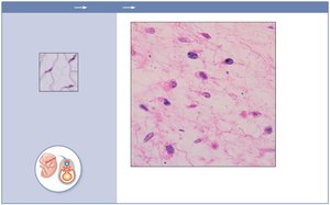
Comparison of Classes of Connective Tissues
The following table summarizes the main subclasses, cell types, matrix composition, and general features of the four major classes of connective tissue.
Tissue Class and Example | Subclasses | Cells | Matrix | General Features |
|---|---|---|---|---|
Connective Tissue Proper | Loose connective (areolar, adipose, reticular); Dense connective (regular, irregular, elastic) | Fibroblasts, fibrocytes, defense cells, fat cells | Gel-like ground substance; all three fiber types: collagen, reticular, elastic | Functions as binding tissue; resists mechanical stress, especially tension |
Cartilage | Hyaline, elastic, fibrocartilage | Chondroblasts (growing cartilage), chondrocytes | Gel-like ground substance; fibers: collagen, elastic (in some) | Resists compression due to large amounts of water in matrix; functions to cushion and support body structures |
Bone Tissue | Compact bone, spongy bone | Osteoblasts, osteocytes | Gel-like ground substance calcified with inorganic salts; fibers: collagen | Hard tissue that resists both compression and tension; functions in support |
Blood | Blood cell formation and differentiation are unique to this tissue | Erythrocytes (RBCs), leukocytes (WBCs), platelets | Plasma; no fibers | A fluid tissue; functions to carry O2, CO2, nutrients, wastes, and other substances |
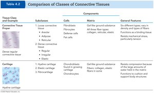
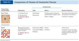
Additional info: The above table is a synthesis of the two-part Table 4.2, summarizing the main features of connective tissue classes for clarity and exam preparation.
