 Back
BackUrinary System: Structure and Function
Study Guide - Smart Notes
 Tailored notes based on your materials, expanded with key definitions, examples, and context.
Urinary System Overview
Functions of the Urinary System
The urinary system is essential for maintaining homeostasis by regulating the volume and composition of blood, removing waste products, and controlling blood pressure. Its three major functions are:
Excretion: Removal of organic waste products from body fluids.
Elimination: Discharge of waste products into the environment.
Homeostatic Regulation: Regulation of blood volume, solute concentrations, and blood pH.
Additional functions include:
Regulating blood volume and blood pressure by adjusting water loss and releasing hormones (erythropoietin and renin).
Regulating plasma concentrations of sodium, potassium, chloride, and other ions.
Stabilizing blood pH by controlling the loss of hydrogen and bicarbonate ions.
Conserving valuable nutrients while excreting waste products such as urea and uric acid.
Assisting the liver in detoxifying poisons.
Organs of the Urinary System
The urinary system consists of the kidneys, ureters, urinary bladder, and urethra.
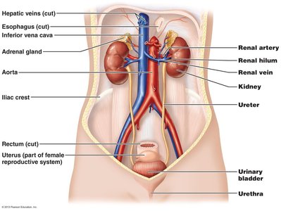
Kidney Anatomy
Regions of the Kidney
The kidney is divided into three main regions:
Cortex: The outer region.
Medulla: The inner region.
Pelvis: The central collecting area.
Blood and Nerve Supply
Blood enters the kidney via the renal artery and leaves through the renal vein. The renal plexus provides nerve supply to the kidney and ureter.
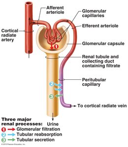
Nephron Structure
The nephron is the functional unit of the kidney, responsible for filtration, reabsorption, and secretion.
Renal Corpuscle: Consists of the glomerulus and Bowman's capsule. Filtration occurs here as blood pressure forces water and solutes out of the glomerulus into the capsular space.
Renal Tubule: Includes the proximal convoluted tubule (PCT), loop of Henle, and distal convoluted tubule (DCT). Responsible for reabsorbing useful substances, water, and secreting waste products.
Proximal Convoluted Tubule (PCT)
Located in the cortex.
Primary function is reabsorption of nutrients and water.
Loop of Henle
Extends into medulla and cortex.
Descending limb is permeable to water; ascending limb pumps sodium and chloride ions out of filtrate.
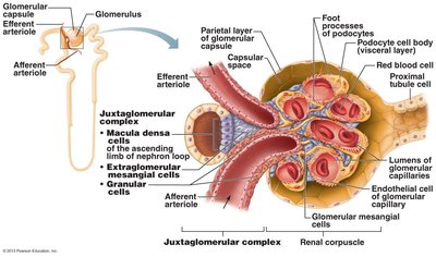
Distal Convoluted Tubule (DCT) and Juxtaglomerular Apparatus (JGA)
DCT is important for secretion and selective reabsorption of ions and water.
JGA is an endocrine structure that secretes erythropoietin and renin, regulating blood pressure and filtration rate.
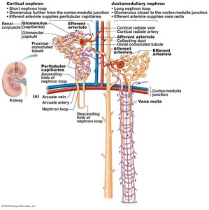
Types of Nephrons
Cortical Nephrons: 85% of nephrons, mostly in cortex.
Juxtamedullary Nephrons: Located near cortex-medulla junction, loops extend deep into medulla, important for producing concentrated urine.
Capillary Beds Associated with Nephrons
Glomerulus: Specialized for filtration; high blood pressure forces fluid and solutes out.
Peritubular Capillaries: Adapted for absorption; low-pressure, porous capillaries absorb solutes and water from tubule cells.
Vasa Recta: Straight vessels associated with juxtamedullary nephrons, important for forming concentrated urine.
Collecting System
DCT opens into the collecting system, which consists of collecting ducts and papillary ducts.
Transports tubular fluid to the renal pelvis and adjusts urine composition and concentration.
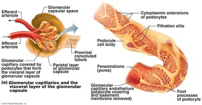
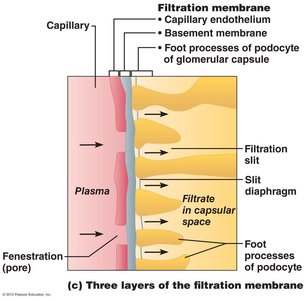
Filtration Membrane
Structure and Function
The filtration membrane lies between the blood and the interior of the glomerular capsule. It is a porous membrane that allows passage of water and small solutes but restricts larger molecules.
Three layers:
Fenestrated endothelium of glomerular capillaries
Visceral membrane of glomerular capsule (podocytes)
Fused basement membrane
Fenestrations allow plasma components but not blood cells.
Basement membrane restricts passage of all but the smallest proteins.
Podocytes engulf and degrade macromolecules stuck in the membrane.
Renal Physiology
Goal of Urine Production
The main goal is to maintain homeostasis by regulating blood volume and composition, and excreting metabolic waste products:
Urea: From amino acid breakdown.
Creatinine: From muscle metabolism.
Uric Acid: From recycling nitrogenous bases.
Basic Processes of Urine Formation
Filtration: Blood pressure forces water and solutes across glomerular capillaries into the capsular space.
Reabsorption: Removal of water and solutes from filtrate into peritubular fluid; selective process involving diffusion and carrier proteins.
Secretion: Transport of solutes from peritubular fluid into tubular fluid; necessary to lower plasma concentration of undesirable materials.
Mechanisms of Transport
Facilitated Diffusion: Carrier protein transports molecules without energy.
Active Transport: Uses energy to move substances against concentration gradient.
Cotransport: Two substances cross membrane together, following concentration gradient of one.
Countertransport: Two ions move in opposite directions across the membrane.
Carrier-Mediated Transport Characteristics
Specific substrate binds to carrier protein.
Carrier proteins usually work in one direction.
Membrane contains many types of carrier proteins.
Carrier proteins can be saturated; transport maximum (Tm) is the concentration at saturation.
Tm and Renal Threshold
Plasma proteins and nutrients are removed from tubular fluid by cotransport or facilitated diffusion.
When concentration exceeds Tm, excess remains in tubular fluid and is excreted in urine.
Overview of Renal Function
Part of Nephron | Function |
|---|---|
Renal Corpuscle (Glomerulus) | Filtration of water and dissolved substances from plasma |
PCT | Reabsorption of glucose, active transport of acids and ions, reabsorption of water and Cl-, active secretion of penicillin, histamine, and H+ |
Descending limb of Loop | Reabsorption of water by osmosis |
Ascending limb of Loop | Reabsorption of sodium, potassium, and Cl- by active transport |
DCT | Reabsorption of sodium ions and water, secretion of H+ and K+ |
Collecting duct | Reabsorption of water by osmosis |
Tubular Secretion
Importance of Secretion
Disposes substances bound to plasma proteins (e.g., drugs, metabolites).
Eliminates undesirable substances not reabsorbed passively (e.g., urea, uric acid).
Eliminates excess potassium.
Controls blood pH by secreting H+ and retaining HCO3-.
Regulation of Filtration Rate
Sympathetic Nervous System
Responds to changes in blood pressure and volume.
Decreased BP/BV causes vasoconstriction of afferent arterioles, reducing GFR and conserving water.
Increased BP/BV causes vasodilation, increasing GFR and urine formation.
Renin-Angiotensin-Aldosterone System
Juxtaglomerular cells secrete renin in response to low BP, sympathetic stimulation, or decreased ion concentration at DCT.
Renin converts angiotensinogen to angiotensin I, then to angiotensin II.
Angiotensin II increases glomerular pressure, stimulates aldosterone secretion, and promotes sodium and water reabsorption.
Sodium and Water Reabsorption
Water reabsorption is closely linked to sodium reabsorption.
Increased sodium reabsorption leads to increased water reabsorption.
Aldosterone stimulates DCT to reabsorb sodium and secrete potassium.
ADH increases permeability of DCT and collecting duct to water, promoting water reabsorption.
Low ADH results in dilute urine and excretion of excess water.
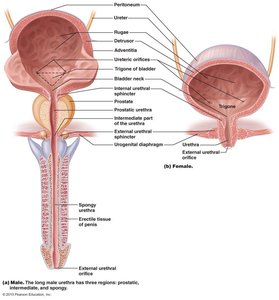
Urine Elimination
Pathway of Urine
Kidneys → Ureters → Urinary Bladder → Urethra → External Environment
Ureters use peristaltic waves to propel urine to the bladder.
Bladder Structure and Micturition Reflex
Bladder has three layers: mucosa, submucosa, and muscularis (detrusor muscle).
Micturition reflex involves contraction of detrusor muscle and relaxation of external urethral sphincter.
Stimulus: Bladder distension activates stretch receptors, triggering reflex center in spinal cord.
External urethral sphincter is skeletal muscle and under conscious control.
Summary Table: Major Processes in the Nephron
Process | Location | Main Function |
|---|---|---|
Filtration | Glomerulus | Removal of water and solutes from blood |
Reabsorption | PCT, Loop of Henle, DCT, Collecting Duct | Recovery of useful substances and water |
Secretion | PCT, DCT | Removal of additional waste and regulation of pH |
Key Equations
Glomerular Filtration Rate (GFR): where is the filtration coefficient, is glomerular capillary hydrostatic pressure, is Bowman's space hydrostatic pressure, and is glomerular capillary oncotic pressure.
Transport Maximum (Tm):
Additional info: Carrier-mediated transport, renal threshold, and hormonal regulation are critical for understanding kidney function and urine formation.
