 Back
BackCell Structure, Membranes, and Transport: Foundational Biochemistry Study Notes
Study Guide - Smart Notes
 Tailored notes based on your materials, expanded with key definitions, examples, and context.
Cell Structure and Function
Introduction to Cells
Cells are the basic functional units of structure and function in all living organisms. They are responsible for carrying out essential biochemical processes necessary for life.
Definition: A cell is the smallest unit of life that can function independently.
Discovery: Cells were discovered by the use of microscopes. Robert Hooke observed cell walls in dead cork, while Anton van Leeuwenhoek observed living cells.
Microscopic Nature: Most cells are too small to be seen with the naked eye.
Cell Theory
Cell theory is a fundamental concept in biology and biochemistry, describing the properties and functions of cells.
All organisms are composed of one or more cells.
The cell is the basic unit of structure and function in organisms.
All cells arise from pre-existing cells.
Cells contain hereditary information (DNA) passed from cell to cell during division.
Prokaryotic vs. Eukaryotic Cells
Cells can be classified as prokaryotic or eukaryotic based on their structural features.
Prokaryotic Cells: Lack a nucleus and membrane-bound organelles. Example: Bacteria.
Eukaryotic Cells: Have a nucleus and membrane-bound organelles. Example: Plants, animals, fungi.
Cell Membranes and Permeability
Structure of Cell Membrane
The cell membrane is a selective barrier that controls the movement of substances in and out of the cell. It is composed mainly of a phospholipid bilayer with embedded proteins.
Phospholipid Bilayer: Consists of two layers of phospholipids with hydrophilic heads facing outward and hydrophobic tails facing inward.
Cholesterol: Present in animal cell membranes, helps maintain fluidity.
Proteins: Integral and peripheral proteins serve various functions such as transport, signaling, and structural support.
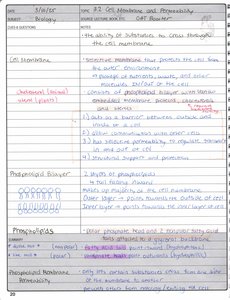
Membrane Permeability
Membrane permeability refers to the ability of substances to cross the cell membrane. It depends on the size, charge, and solubility of molecules.
Small, nonpolar molecules (e.g., O2, CO2) can diffuse freely.
Large or charged molecules require transport proteins.
Water moves via osmosis through aquaporins.
Organelles and Cell Structures
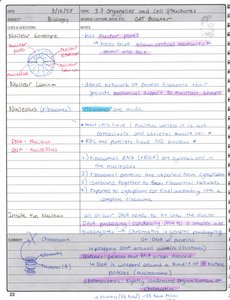
Nucleus
The nucleus is the control center of the cell, containing genetic material (DNA).
Nuclear Envelope: Double membrane that surrounds the nucleus.
Nucleolus: Site of ribosome synthesis.
Nuclear Lamina: Network of proteins providing structural support.
Cytoplasm and Cytosol
The cytoplasm includes all contents within the cell membrane except the nucleus. The cytosol is the fluid portion.
Cytoplasm: Contains organelles and cytosol.
Cytosol: Jelly-like fluid where metabolic reactions occur.
Ribosomes
Ribosomes are the sites of protein synthesis.
Free Ribosomes: Located in cytosol, synthesize proteins for use within the cell.
Bound Ribosomes: Attached to endoplasmic reticulum, synthesize proteins for secretion or membrane insertion.
Endoplasmic Reticulum (ER)
The ER is a network of membranes involved in protein and lipid synthesis.
Rough ER: Studded with ribosomes, synthesizes proteins.
Smooth ER: Lacks ribosomes, synthesizes lipids and detoxifies chemicals.
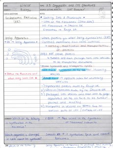
Golgi Apparatus
The Golgi apparatus modifies, sorts, and packages proteins and lipids for secretion or delivery to other organelles.
Receives proteins from ER.
Processes and packages proteins into vesicles.
Lysosomes and Peroxisomes
Lysosomes contain digestive enzymes for breaking down waste, while peroxisomes detoxify harmful substances.
Lysosomes: Digest cellular waste and pathogens.
Peroxisomes: Break down fatty acids and detoxify hydrogen peroxide.
Mitochondria
Mitochondria are the powerhouses of the cell, generating ATP through cellular respiration.
Structure: Double membrane, inner membrane forms cristae.
Function: Site of aerobic respiration and energy production.
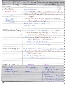
Chloroplasts (Plant Cells Only)
Chloroplasts are organelles found in plant cells responsible for photosynthesis.
Contain chlorophyll, capture light energy.
Convert CO2 and H2O into glucose and O2.
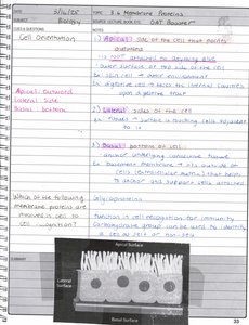
Membrane Proteins
Classes of Membrane Proteins
Membrane proteins are classified based on their association with the lipid bilayer.
Peripheral Proteins: Not embedded, attached to membrane surface.
Integral Proteins: Embedded within the membrane, often span the bilayer.
Transmembrane Proteins: Span the entire membrane, involved in transport and signaling.
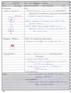
Protein Functions
Carrier Proteins: Transport specific substances across the membrane.
Receptor Proteins: Receive and transmit signals.
Glycoproteins: Proteins with carbohydrate chains, important for cell recognition.
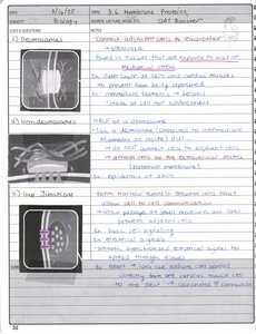
Membrane Junctions
Types of Junctions
Cells are connected by specialized junctions that facilitate communication and adhesion.
Tight Junctions: Seal adjacent cells, prevent leakage.
Desmosomes: Anchor cells together, provide mechanical strength.
Gap Junctions: Allow passage of ions and small molecules between cells.
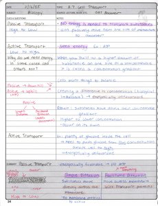
Cell Transport
Passive Transport
Passive transport involves the movement of substances across membranes without energy input.
Diffusion: Movement from high to low concentration.
Osmosis: Diffusion of water across a membrane.
Facilitated Diffusion: Uses transport proteins for movement of larger or charged molecules.
Active Transport
Active transport requires energy (usually ATP) to move substances against their concentration gradient.
Primary Active Transport: Direct use of ATP (e.g., sodium-potassium pump).
Secondary Active Transport: Uses energy from another gradient.
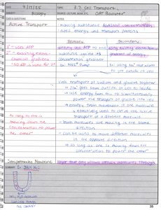
Endocytosis and Exocytosis
Cells transport large molecules via vesicles in processes called endocytosis (into cell) and exocytosis (out of cell).
Phagocytosis: Cell engulfs large particles.
Pinocytosis: Cell takes in fluid.
Receptor-mediated Endocytosis: Specific uptake of molecules.
Osmosis and Tonicity
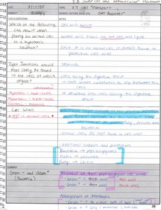
Osmosis affects cell volume depending on the tonicity of the surrounding solution.
Hypertonic: Higher solute concentration outside, cell shrinks.
Hypotonic: Lower solute concentration outside, cell swells.
Isotonic: Equal solute concentration, cell remains unchanged.
Extracellular Matrix and Cell Movement
Extracellular Matrix (ECM)
The ECM provides structural support and mediates cell signaling.
Components: Glycoproteins, proteoglycans, collagen, fibronectin.
Functions: Structural support, cell adhesion, communication.
Cell Movement
Cells move using cytoskeletal elements such as microtubules, microfilaments, and motor proteins.
Microtubules: Provide tracks for organelle movement.
Microfilaments: Involved in cell shape and movement.
Motor Proteins: Kinesin and dynein move along microtubules.
Summary Table: Prokaryotic vs. Eukaryotic Cells
Feature | Prokaryotic Cells | Eukaryotic Cells |
|---|---|---|
Nucleus | No | Yes |
Membrane-bound Organelles | No | Yes |
Cell Wall | Yes (most) | Some (plants, fungi) |
Size | Small (1-10 µm) | Larger (10-100 µm) |
Examples | Bacteria, Archaea | Plants, Animals, Fungi |
Summary Table: Membrane Transport Types
Transport Type | Energy Required | Direction | Example |
|---|---|---|---|
Passive Transport | No | High to Low | Diffusion, Osmosis |
Facilitated Diffusion | No | High to Low | Glucose transport |
Active Transport | Yes (ATP) | Low to High | Sodium-potassium pump |
Endocytosis | Yes | Into cell | Phagocytosis |
Exocytosis | Yes | Out of cell | Secretion of hormones |
Key Equations
Osmosis: Water movement across a membrane is driven by differences in solute concentration.
Diffusion Rate: Fick's Law describes the rate of diffusion:
where J is the flux, D is the diffusion coefficient, and dC/dx is the concentration gradient.
Active Transport: Energy requirement for moving ions against gradient:
where R is the gas constant, T is temperature, z is ion charge, F is Faraday's constant, and \Delta \Psi is membrane potential.
