 Back
BackA Tour of the Cell & Membrane Structure and Function
Study Guide - Smart Notes
 Tailored notes based on your materials, expanded with key definitions, examples, and context.
Chapter 6: A Tour of the Cell
Cell Structure and Function
Cells are the fundamental units of life, and their structure is closely related to their function. Understanding the differences between cell types and their organelles is essential for grasping how life operates at the microscopic level.
Structure relates to function: The shape and organization of a cell and its components enable it to perform specific tasks efficiently.
Microscopy and Cell Study
Light Microscopes: Allow observation of living cells but cannot resolve small organelles. Image quality depends on magnification, resolution, and contrast.
Scanning Electron Microscopes (SEM): Provide detailed 3D images of cell surfaces.
Transmission Electron Microscopes (TEM): Reveal internal structures of cells.
Cell Fractionation
Cell fractionation is a laboratory technique used to separate organelles from cells, allowing for the study of individual cell components.
Prokaryotic vs. Eukaryotic Cells
Prokaryotic Cells: Found in bacteria and archaea. Characterized by the absence of a nucleus and membrane-bound organelles. DNA is located in the nucleoid region. These cells are generally smaller and simpler than eukaryotic cells.
Eukaryotic Cells: Found in plants, animals, fungi, and protists. Characterized by the presence of a nucleus and membrane-bound organelles. These cells are larger and more complex.
Basic Features of All Cells
Plasma membrane
Cytoplasm
DNA (genetic material/chromosomes)
Ribosomes
Plasma Membrane Structure
The plasma membrane is a selectively permeable barrier composed of a phospholipid bilayer with embedded proteins, cholesterol, and carbohydrates. It regulates the movement of substances into and out of the cell.
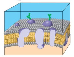
Phospholipid bilayer: Hydrophilic heads face outward, hydrophobic tails face inward.
Membrane proteins: Include channels and transport proteins.
Cholesterol: Stabilizes membrane fluidity.
Carbohydrate chains: Involved in cell recognition.
Surface Area to Volume Ratio
The efficiency of a cell depends on its surface area-to-volume ratio. A higher ratio allows for more efficient exchange of materials, while a lower ratio can limit cell function.
Too little surface area: Cell becomes inefficient at exchanging materials.
Too much surface area: Not efficient for storage or normal function.
Ideal: Maximizes exchange efficiency for cell survival.
Animal and Plant Cell Structure
Animal and plant cells share many organelles but also have unique structures. Understanding these differences is key to recognizing their specialized functions.
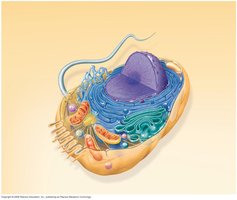
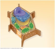
Nucleus: Stores DNA and proteins.
Nuclear envelope: Double membrane with pores surrounding the nucleus.
Chromatin: Loose DNA and proteins.
Chromosomes: Condensed, tightly packed DNA.
Nucleolus: Produces rRNA and assembles ribosomes.
Ribosomes
Free ribosomes: Float in cytosol; synthesize proteins for use within the cell.
Bound ribosomes: Attached to rough ER; synthesize proteins for secretion or membrane insertion.
Endomembrane System
Nuclear envelope
Endoplasmic reticulum (smooth and rough)
Golgi apparatus
Vesicles/vacuoles
Plasma membrane
Lysosomes
Endoplasmic Reticulum (ER)
Smooth ER: Synthesizes lipids, detoxifies drugs/poisons, stores calcium ions, and metabolizes carbohydrates.
Rough ER: Studded with ribosomes; synthesizes proteins for secretion and membrane production.
Golgi Apparatus
Composed of flattened sacs called cisternae.
Modifies, stores, and packages proteins into vesicles for transport.
Lysosomes
Digest and break down food, waste, and old organelles.
Phagocytosis
Process by which a cell engulfs large particles ('cell eating').
Vacuoles
Food vacuole: Stores food and aids in digestion.
Contractile vacuole: Pumps out excess water (common in protists).
Central vacuole: Found in plant cells; stores water and helps maintain cell structure.
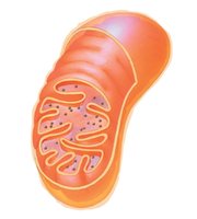
Mitochondria and Chloroplasts
Both mitochondria and chloroplasts are energy-converting organelles with their own DNA and ribosomes, supporting the endosymbiotic theory of their origin.
Mitochondria: Site of cellular respiration; contains outer membrane, inner membrane, cristae (folds), and matrix (fluid).
Chloroplasts: Site of photosynthesis in plants; contains thylakoids (membrane sacs), granum (stack of thylakoids), and stroma (fluid).
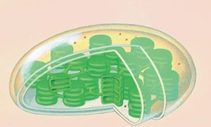
Common features: Double membrane, own DNA, ribosomes, energy production.
Peroxisomes
Break down fatty acids and detoxify harmful substances.
Cytoskeleton
Microtubules: Provide shape, serve as tracks for movement, and are involved in chromosome movement (e.g., cilia, flagella, spindle fibers).
Microfilaments: Maintain cell shape, enable muscle movement, and are involved in cell division (e.g., cell crawling, contraction, cytokinesis).
Intermediate filaments: Provide strength and support, anchor the nucleus, and stabilize organelles.
Motor Proteins
Move vesicles and organelles along microtubules.
Centrosomes and Centrioles
Centrosome: Microtubule-organizing center.
Centrioles: Two cylinders within the centrosome (found in animal cells).
Cilia, Flagella, and Amoeboid Movement
Cilia: Short, numerous structures that move the cell or fluid across the cell surface.
Flagella: Long, few in number; move the cell.
Amoeboid movement: Cell crawling using pseudopodia ('false feet').
Cytoplasmic Streaming
Flow of cytoplasm within a cell to distribute materials.
Extracellular Structures
Cell walls (plants): Provide structure and support; consist of primary and secondary layers.
Extracellular matrix (ECM, animals): Supports, adheres, enables movement, and facilitates cell signaling. Integrins connect ECM to the cytoskeleton.
Intercellular junctions: Include plasmodesmata (plants), tight junctions, desmosomes, and gap junctions (animals).
Chapter 7: Membrane Structure and Function
Selective Permeability
The plasma membrane allows some substances to cross more easily than others, maintaining the internal environment of the cell.
Phospholipids and the Fluid Mosaic Model
Amphipathic molecules: Phospholipids have hydrophilic (polar) heads and hydrophobic (nonpolar) tails.
Fluid mosaic model: Describes the dynamic arrangement of phospholipids and proteins in the membrane.
Phospholipid movement: Lateral movement is common; flip-flop between layers is rare.
Unsaturated fatty acids: Have double bonds, kinks, and increase membrane fluidity.
Saturated fatty acids: No double bonds, straight chains, decrease fluidity.
Membrane Proteins
Peripheral proteins: Loosely bound to the membrane surface.
Integral proteins: Embedded in the membrane; may span the bilayer.
N-terminus: Amino end of a protein (–NH2 group).
C-terminus: Carboxyl end of a protein (–COOH group).
Functions of Membrane Proteins
Transport
Enzymatic activity
Signal transduction (receptors)
Cell-cell recognition
Intercellular joining
Attachment to cytoskeleton and ECM
Cell Recognition
Carbohydrates on the membrane surface enable cells to recognize each other.
Vesicle Transport Pathway
Proteins travel from the rough ER to the Golgi apparatus, then are packaged into vesicles and transported to the plasma membrane.
Transport Proteins
Aquaporins: Channel proteins for water transport.
Carrier proteins: Change shape to move molecules across the membrane.
Diffusion and Osmosis
Diffusion: Movement of molecules from high to low concentration (down the concentration gradient).
Equilibrium: State where molecules are evenly distributed.
Osmosis: Diffusion of water across a membrane.
Tonicity and Water Balance
Isotonic solution: Equal solute concentration; no net water movement.
Hypertonic solution: Higher solute concentration outside; water leaves the cell.
Hypotonic solution: Lower solute concentration outside; water enters the cell.
Osmoregulation: Regulation of water balance in cells.
Plant cells: Turgid (firm, full of water), flaccid (limp), plasmolysis (membrane pulls away from wall).
Facilitated Diffusion
Passive transport using channel or carrier proteins (e.g., aquaporins for water, ion channels for ions).
Active Transport
Requires ATP to move molecules against their concentration gradient.
Sodium-potassium pump: Uses ATP to pump Na+ out and K+ in.
Membrane Potential and Electrochemical Gradients
Membrane potential: Voltage difference across the membrane.
Electrochemical gradient: Combination of ion concentration and charge difference.
Electrogenic pump: Generates voltage across the membrane (e.g., sodium-potassium pump, proton pump).
Bulk Transport: Endocytosis and Exocytosis
Phagocytosis: 'Cell eating'—engulfing large particles into vesicles.
Pinocytosis: 'Cell drinking'—taking in extracellular fluid and dissolved substances in small vesicles.
