 Back
A Tour of the Cell: Microscopy, Cell Structure, and Function Part 1
Study Guide - Smart Notes
 Tailored notes based on your materials, expanded with key definitions, examples, and context.
Concept 6.1: Biologists Use Microscopes and Biochemistry to Study Cells
Microscopy in Cell Biology
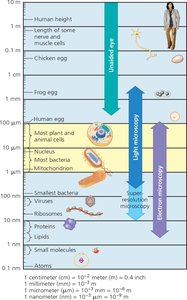
Microscopy is essential for visualizing cells, which are typically too small to be seen by the naked eye. Different types of microscopes allow biologists to study cell structure and function at various levels of detail.
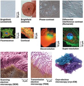
Light Microscopy (LM): Uses visible light and glass lenses to magnify images of specimens. Suitable for observing live cells and general cell structure.
Electron Microscopy (EM): Uses electron beams with much shorter wavelengths than light, providing much higher resolution. Includes Scanning Electron Microscopes (SEM) for surface details and Transmission Electron Microscopes (TEM) for internal structures. Preparation for EM kills cells.
Advanced Light Microscopy: Techniques such as confocal and deconvolution microscopy produce sharper, three-dimensional images of tissues and cells.
Key Parameters of Microscopy:
Magnification: Ratio of image size to actual size (up to ~1,000× for LM).
Resolution: Clarity of the image; minimum distance between two distinguishable points. Resolution improves with shorter wavelengths.
Contrast: Visible differences in brightness; enhanced by staining or labeling.
Limitations: Light microscopes cannot resolve most organelles; electron microscopes require non-living samples.
Example: To observe live cells, use a light microscope. To study the internal structure of organelles, use a TEM.
Concept 6.2: Eukaryotic Cells Have Internal Membranes That Compartmentalize Their Functions
Prokaryotic vs. Eukaryotic Cells
All living organisms are composed of either prokaryotic or eukaryotic cells. The main distinction is the presence of membrane-bound organelles, especially the nucleus, in eukaryotes.
Prokaryotic Cells: Found in Bacteria and Archaea. DNA is located in the nucleoid, not enclosed by a membrane. Lack membrane-bound organelles.
Eukaryotic Cells: Found in protists, fungi, animals, and plants. DNA is enclosed within a double-membrane nucleus. Contain various membrane-bound organelles.
Common Features: All cells have a plasma membrane, cytosol, chromosomes (DNA), and ribosomes.
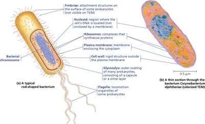
Cytoplasm: In prokaryotes, the entire interior; in eukaryotes, the region between the nucleus and plasma membrane.
Cell Size: Eukaryotic cells are generally larger. Surface area-to-volume ratio limits cell size; as cells grow, volume increases faster than surface area.
Biological Membranes
Membranes are primarily composed of a phospholipid bilayer with embedded proteins and other lipids. Each membrane's composition reflects its specific function.
Phospholipid Bilayer: Provides the basic structure of all biological membranes.
Proteins: Embedded or attached; responsible for specific functions such as transport, signaling, and enzymatic activity.
Membrane Diversity: Different organelles have unique membrane compositions suited to their roles (e.g., mitochondrial membranes contain enzymes for cellular respiration).
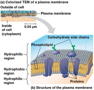
Example: The plasma membrane regulates the exchange of materials, maintaining the internal environment of the cell.
Animal and Plant Cells: A Panoramic View
Both animal and plant cells are eukaryotic and share most organelles, but plant cells have unique structures such as cell walls, chloroplasts, and large central vacuoles.
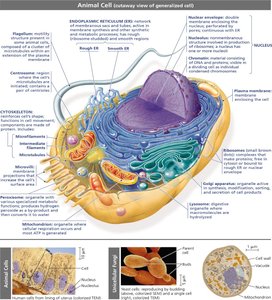
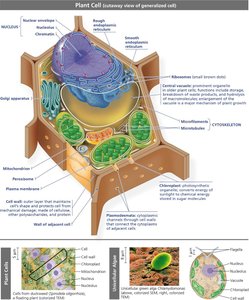
Example: Animal cells lack cell walls and chloroplasts, while plant cells contain both for structural support and photosynthesis, respectively.
Concept 6.3: The Eukaryotic Cell’s Genetic Instructions Are Housed in the Nucleus and Carried Out by the Ribosomes
The Nucleus: Information Central
The nucleus is the control center of the eukaryotic cell, containing most of the cell’s genetic material and coordinating activities such as gene expression and DNA replication.
Nuclear Envelope: Double membrane that encloses the nucleus, separating it from the cytoplasm. Contains nuclear pores for regulated exchange of materials.
Nuclear Lamina: Protein network lining the inner membrane, maintaining nuclear shape.
Nuclear Matrix: Internal framework providing structural support and organization.
Chromosomes: DNA molecules associated with histone proteins, forming chromatin. Each chromosome is a single DNA molecule with associated proteins.
Nucleolus: Dense region where ribosomal RNA (rRNA) is synthesized and ribosomal subunits are assembled.
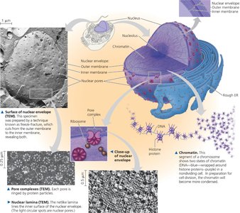
Example: Ribosomal subunits are produced in the nucleolus and exported to the cytoplasm for protein synthesis.
Ribosomes: Protein Factories
Ribosomes are complexes of rRNA and proteins that carry out protein synthesis. They can be free in the cytosol or bound to the endoplasmic reticulum (ER) or nuclear envelope.
Free Ribosomes: Synthesize proteins that function in the cytosol.
Bound Ribosomes: Synthesize proteins for insertion into membranes, packaging into organelles, or export from the cell.
Cells with High Protein Production: Such as pancreatic cells, have many ribosomes and prominent nucleoli.
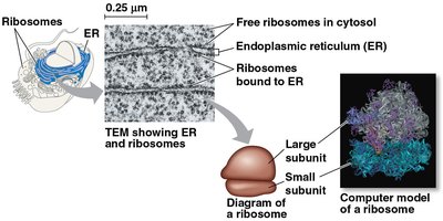
Example: Digestive enzymes secreted by pancreatic cells are synthesized by bound ribosomes on the ER.
Summary Table: Comparison of Prokaryotic and Eukaryotic Cells
Feature | Prokaryotic Cells | Eukaryotic Cells |
|---|---|---|
DNA Location | Nucleoid (no membrane) | Nucleus (double membrane) |
Membrane-bound Organelles | Absent | Present |
Cell Size | Generally smaller | Generally larger |
Domains | Bacteria, Archaea | Protists, Fungi, Plants, Animals |
Key Terms and Definitions
Magnification:
Resolution: Minimum distance between two distinguishable points.
Phospholipid Bilayer: Double layer of phospholipids forming the basic structure of cell membranes.
Chromatin: Complex of DNA and proteins forming chromosomes.
Ribosome: Cellular structure that synthesizes proteins from mRNA templates.
