 Back
BackA Tour of the Cell: Structure, Function, and Diversity
Study Guide - Smart Notes
 Tailored notes based on your materials, expanded with key definitions, examples, and context.
A Tour of the Cell
Introduction to Cell Theory
Cells are the fundamental units of life, forming the basis for all living organisms. The cell theory provides the foundation for understanding biological structure and function.
All organisms are composed of cells.
The cell is the basic unit of structure and organization in organisms.
All cells arise from pre-existing cells.
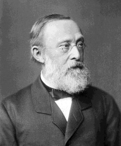
Additional info: Rudolf Virchow contributed the concept "Omnis cellula e cellula" (all cells come from cells), emphasizing the continuity of life.
Unifying Features of All Cells
Despite their diversity, all cells share certain structural and functional characteristics:
Plasma membrane: A selective barrier that regulates the passage of substances.
Cytoplasm: The semifluid substance within the membrane containing organelles and molecules.
Genetic material: DNA serves as the hereditary blueprint.
Ribosomes: Sites of protein synthesis.
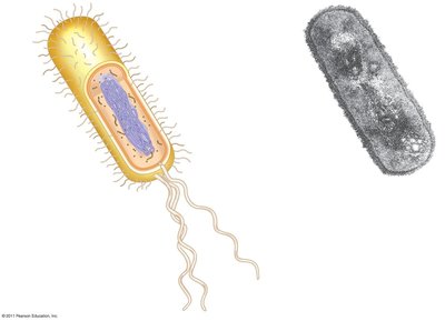
Prokaryotes vs. Eukaryotes
Major Differences
Cells are classified as either prokaryotic or eukaryotic based on structural differences.
Prokaryotic cells: Lack a nucleus and membrane-bound organelles; DNA is located in the nucleoid region.
Eukaryotic cells: Possess a true nucleus and numerous membrane-bound organelles.
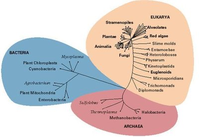
Additional info: The three domains of life—Bacteria, Archaea, and Eukarya—reflect evolutionary relationships among cell types.
Structural Comparison
Feature | Eukaryotes | Prokaryotes |
|---|---|---|
Plasma membrane | Yes | Yes |
Cytoplasm | Yes | Yes |
Nucleus | Yes | No |
Nucleoid | No | Yes |
Ribosomes | Yes | Yes |
Membrane-bound organelles | Yes | No |
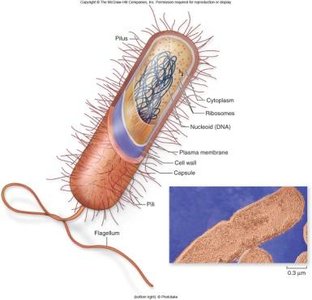
Prokaryotic Cell Structure
Cell wall: Provides structural support and shape.
Capsule: Offers protection and helps cells adhere to surfaces.
Fimbriae and flagella: Aid in attachment and movement.
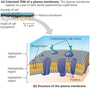
Plasma Membrane Structure
The plasma membrane is a phospholipid bilayer with embedded proteins, crucial for maintaining cellular homeostasis.
Hydrophilic heads face outward; hydrophobic tails face inward.
Proteins serve as channels, receptors, and enzymes.
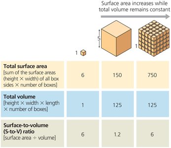
Surface Area to Volume Ratio
Cell size is limited by the ratio of surface area to volume, which affects the efficiency of material exchange.
As cells grow, volume increases faster than surface area.
Small cells have a higher surface area-to-volume ratio, facilitating efficient transport.
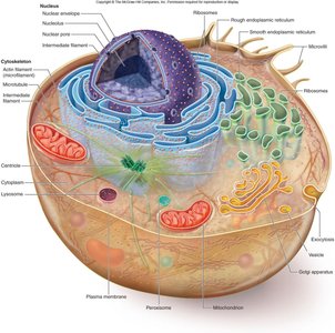
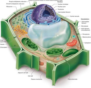
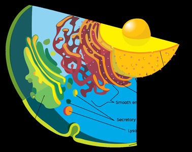
Eukaryotic Cell Compartmentalization
Internal Membranes and Organelles
Eukaryotic cells contain internal membranes that compartmentalize functions, allowing for specialization and efficiency.
Nucleus: Stores genetic information.
Endoplasmic reticulum (ER): Synthesizes proteins and lipids.
Golgi apparatus: Modifies, sorts, and packages proteins and lipids.
Lysosomes and vacuoles: Involved in digestion and storage.
Mitochondria and chloroplasts: Sites of energy transformation.

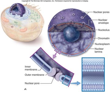
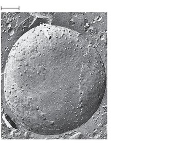
The Nucleus: Information Central
Structure and Function
The nucleus is the control center of the cell, containing most of the cell's genetic material.
Nuclear envelope: Double membrane with pores for molecular transport.
Chromatin: DNA-protein complex that condenses to form chromosomes during cell division.
Nucleolus: Site of ribosomal RNA (rRNA) synthesis and ribosome assembly.
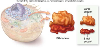
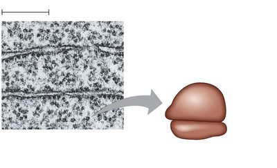
Ribosomes: Protein Factories
Structure and Function
Ribosomes are complexes of rRNA and proteins that carry out protein synthesis in the cytoplasm or on the endoplasmic reticulum.
Free ribosomes: Synthesize proteins for use within the cytosol.
Bound ribosomes: Attached to the rough ER, synthesize proteins for membranes or secretion.

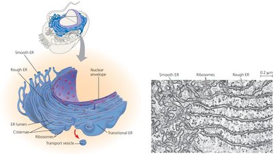
The Endomembrane System
Components and Functions
The endomembrane system is a network of membranes involved in protein and lipid synthesis, modification, and transport.
Nuclear envelope
Endoplasmic reticulum (ER): Smooth and rough regions with distinct functions.
Golgi apparatus
Lysosomes
Vacuoles
Plasma membrane
Endoplasmic Reticulum (ER)
Smooth ER: Synthesizes lipids, metabolizes carbohydrates, detoxifies drugs, stores calcium ions.
Rough ER: Studded with ribosomes; synthesizes proteins and glycoproteins, distributes transport vesicles, and produces membranes.
Golgi Apparatus
The Golgi apparatus consists of flattened sacs (cisternae) and functions as the cell's shipping and receiving center.
Modifies products from the ER.
Sorts and packages materials into transport vesicles.
Vacuoles
Vacuoles are large vesicles with diverse functions, including storage, waste disposal, and maintaining turgor pressure in plant cells.
Food vacuoles: Formed by phagocytosis.
Contractile vacuoles: Pump excess water out of cells (in protists).
Central vacuole: Found in plant cells, stores ions and helps maintain cell rigidity.
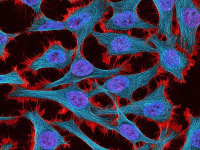
Cytoskeleton and Cell Movement
Overview of the Cytoskeleton
The cytoskeleton is a dynamic network of protein fibers that provides structural support, organizes organelles, and facilitates cell movement.
Microtubules: Hollow rods made of tubulin; involved in cell shape, organelle movement, and chromosome separation.
Microfilaments (Actin filaments): Thin rods that bear tension and enable cell movement.
Intermediate filaments: Provide mechanical support and maintain cell shape.
Microtubules
Shape and support the cell.
Guide movement of organelles and vesicles.
Form the spindle apparatus during cell division.
Microfilaments (Actin Filaments)
Bear tension and resist pulling forces.
Enable cell crawling and muscle contraction (with myosin).
Intermediate Filaments
More permanent structures than microtubules or microfilaments.
Reinforce cell shape and anchor organelles.
Mitochondria and Chloroplasts
Energy Transformation
Mitochondria and chloroplasts are specialized organelles responsible for energy conversion in eukaryotic cells.
Mitochondria: Site of cellular respiration; converts organic molecules into ATP.
Chloroplasts: Site of photosynthesis in plant cells and algae.
