 Back
BackA Tour of the Cell: Structure, Function, and Diversity
Study Guide - Smart Notes
 Tailored notes based on your materials, expanded with key definitions, examples, and context.
Chapter 6: A Tour of the Cell
Introduction to Cell Diversity
Cells are the fundamental units of life, exhibiting remarkable diversity in their structures, macromolecules, and processes. Understanding cell structure and function is essential for grasping the complexity of biological systems.
History of Cell Discovery
Development of Microscopy
Robert Hooke (1665): Used a simple microscope to observe cork, coining the term "cellulae" for the small compartments he saw.
Antonie van Leeuwenhoek (1680s): Improved microscopes and was the first to observe living microorganisms in water.
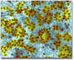
Microscopy: Revolutionized biology by revealing the presence and structure of cells.
Magnification: Ratio of the image size to the actual size of the object.
Resolution: Measure of image clarity; the ability to distinguish two close objects as separate.
Contrast: Difference in brightness between light and dark areas, enhancing visibility of structures.
Microscopy Techniques
Types of Microscopes
Light Microscopes: Use visible light; magnify up to ~1000x.
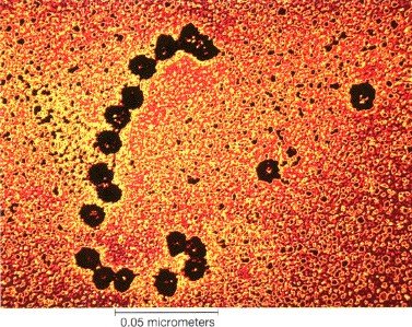
Transmission Electron Microscopes (TEM): Use electrons to visualize internal cell structures; magnify up to ~500,000x.
Scanning Electron Microscopes (SEM): Provide 3D images of cell surfaces.
Cryo-electron Microscopy: Preserves specimens in a near-native state for imaging.
Cell Fractionation: Technique to separate cellular components for study.
Cell Types: Prokaryotic vs. Eukaryotic
Major Differences
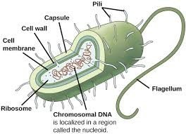
Prokaryotic Cells: Lack a nucleus; DNA is in a nucleoid region. No membrane-bound organelles. Examples: Bacteria and Archaea.
Eukaryotic Cells: Have a true nucleus surrounded by a nuclear envelope and many membrane-bound organelles. Found in animals, plants, fungi, and protists.
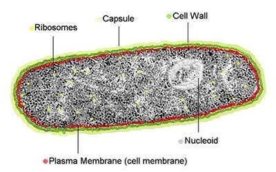
Prokaryotic Cell Structure
Nucleoid: Region containing circular DNA.
Cell Wall: Provides structure and protection.
Capsule/Slime Layer: Offers additional protection and helps in adhesion.
Ribosomes: Sites of protein synthesis.
Lack of Membrane-bound Organelles: No nucleus, mitochondria, or other organelles.
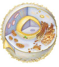
Eukaryotic Cell Structure
Nucleus: Contains chromosomes and separates genetic material from the cytoplasm.
Membrane-bound Organelles: Specialized compartments for various cellular functions.
Examples: Animal, plant, fungal, and protist cells.
Cytoplasm and Organelles
Cytoplasm
Cytoplasm: The region between the plasma membrane and the nucleus; contains organelles suspended in cytosol (watery part).
Function: Site for most cellular activities; acts as the "factory floor" of the cell.
Organelles
Definition: Specialized structures within cells, each with a specific function.
Examples: Nucleus, mitochondria, endoplasmic reticulum, Golgi apparatus, lysosomes, vacuoles.
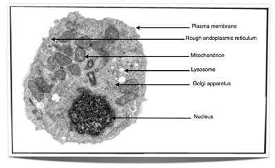
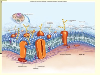
Plasma Membrane
Structure and Function
Phospholipid Bilayer: Forms the basic structure, with hydrophilic heads facing outward and hydrophobic tails inward.
Embedded Proteins: Serve as channels, receptors, and enzymes.
Function: Acts as the "gatekeeper," controlling the movement of substances in and out of the cell.
Nucleus and Genetic Material
Nucleus
Nuclear Envelope: Double membrane continuous with the endoplasmic reticulum; contains nuclear pores for transport.
Function: Houses chromosomes; controls cellular reproduction and protein synthesis.
Nucleolus: Site of ribosome synthesis.
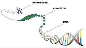
Chromosomes and Chromatin
Chromatin: DNA-protein complex; appears grainy and threadlike except during cell division.
Chromosomes: Condensed chromatin visible during cell division.
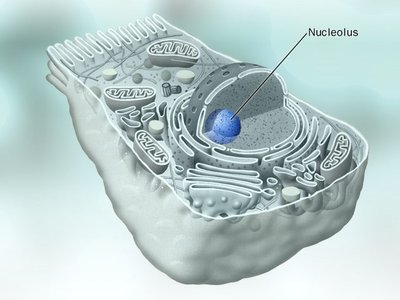
Nucleolus
Structure: Dense region within the nucleus composed of chromatin, RNA, and proteins.
Function: Ribosome assembly.
Ribosomes
Structure and Function
Composition: Made of ribosomal RNA (rRNA) and proteins; not membrane-bound.
Function: Site of protein synthesis.
Location: Free in cytoplasm or attached to the endoplasmic reticulum (ER).
Polyribosomes: Groups of ribosomes translating the same mRNA.
The Endomembrane System
Components and Functions
Includes: Nuclear envelope, endoplasmic reticulum (ER), Golgi apparatus, lysosomes, vesicles, vacuoles, and plasma membrane.
Function: Synthesis, modification, packaging, and transport of proteins and lipids.
Absent in Prokaryotes: Only eukaryotic cells possess this system.
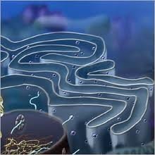
Endoplasmic Reticulum (ER)
Structure: Network of membranous channels and sacs connected to the nuclear envelope.
Function: Synthesis of proteins (rough ER) and lipids (smooth ER).
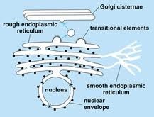
Smooth ER vs. Rough ER
Smooth ER: Lacks ribosomes; synthesizes lipids, detoxifies drugs, stores calcium ions, and metabolizes carbohydrates.
Rough ER: Studded with ribosomes; synthesizes proteins for export, modifies proteins, and forms vesicles for transport to the Golgi apparatus.
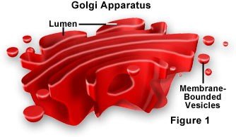
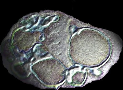
Golgi Apparatus
Structure: Stack of flattened membranous sacs (cisternae).
Function: Processes, packages, and secretes proteins and lipids; forms polysaccharides; cis side receives, trans side ships out.
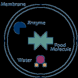
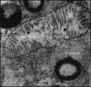
Lysosomes
Structure: Membranous vesicles containing hydrolytic (digestive) enzymes.
Function: Intracellular digestion (autophagy); breakdown of viruses, bacteria, and cellular debris.
Vacuoles
Structure: Membrane-enclosed sacs.
Types: Food vacuoles, contractile vacuoles (in protists), central vacuole (in plants).
Function: Storage of organic compounds, pigments, and water; maintenance of turgor pressure in plants.
Energy-Related Organelles
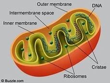
Mitochondria
Function: Site of aerobic cellular respiration; converts food into usable energy (ATP).
Structure: Double membrane; inner membrane forms cristae, matrix is the inner fluid-filled space.
"Powerhouse of the cell": Generates most of the cell's ATP.
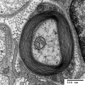
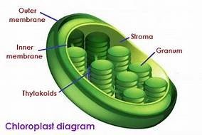
Chloroplasts
Function: Site of photosynthesis; converts light energy into glucose.
Structure: Double membrane; contains stroma (fluid), thylakoids (membranous sacs), and grana (stacks of thylakoids).
Other Plastids: Amyloplasts (store starch), chromoplasts (store pigments).
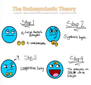
Endosymbiotic Theory
Explanation: Mitochondria and chloroplasts originated as free-living prokaryotes engulfed by ancestral eukaryotic cells.
Evidence: Both organelles contain their own DNA and ribosomes, and replicate independently within the cell.
Other Organelles and Structures
Peroxisomes
Structure: Single membrane-bound organelles.
Function: Break down fatty acids, detoxify alcohol, and convert hydrogen peroxide (H2O2) to water.
Cytoskeleton
Function: Provides cell shape, anchors organelles, and enables movement.
Components: Microtubules (thick), intermediate filaments (medium), microfilaments (thin).
Microtubules
Structure: Hollow tubes made of tubulin.
Function: Maintain cell shape, facilitate vesicle movement, form centrioles, cilia, and flagella.
Microfilaments
Structure: Thin rods made of actin.
Function: Muscle contraction, cell movement (pseudopodia), cytoplasmic streaming in plants.
Intermediate Filaments
Structure: Fibrous proteins; more permanent than microtubules or microfilaments.
Function: Maintain cell shape, anchor nucleus and other organelles.
Cell Wall and Cell Junctions
Cell Wall
Found in: Plants, fungi, and some protists.
Structure: Thick, rigid mesh of cellulose fibers (in plants).
Function: Provides protection and structural support.
Cell Junctions
Plasmodesmata: Channels connecting plant cells, allowing transport and communication.
Tight Junctions: Seal neighboring animal cells together.
Desmosomes: Anchor animal cells together.
Gap Junctions: Allow communication between animal cells.
