 Back
BackA Tour of the Cell: Structure, Function, and Microscopy
Study Guide - Smart Notes
 Tailored notes based on your materials, expanded with key definitions, examples, and context.
The Fundamental Units of Life
Introduction to Cells
Cells are the basic units of life, forming the simplest collection of matter that can be alive. All organisms are composed of cells, which may exist as single cells or as part of multicellular organisms. Despite their diversity, all cells share certain structural features and are studied using microscopy and biochemical techniques.
Cell: The smallest unit of life capable of carrying out all life processes.
Unicellular organisms: Organisms consisting of a single cell.
Multicellular organisms: Organisms composed of many cells with specialized functions.
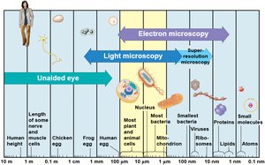
Microscopy
Principles and Types of Microscopy
Microscopes are essential tools for visualizing cells and their components. The three main parameters of microscopy are magnification, resolution, and contrast. Different types of microscopes and techniques are used to study cells at various levels of detail.
Magnification: The ratio of an object's image size to its real size.
Resolution: The clarity of the image, or the minimum distance between two distinguishable points.
Contrast: Visible differences in brightness between parts of the sample.
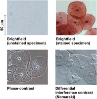
Light Microscopy (LM)
Light microscopes use visible light and glass lenses to magnify images of specimens. They can magnify up to about 1,000 times, but their resolution is limited to about 200 nm, which is insufficient for viewing most organelles.
Various techniques enhance contrast, such as staining and labeling.
Light microscopy allows the study of living cells.
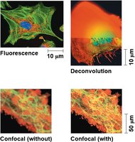
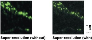
Advanced Light Microscopy Techniques
Recent advances include fluorescence microscopy, confocal microscopy, deconvolution, and super-resolution microscopy, which improve the level of detail and allow visualization of specific cell components.
Electron Microscopy (EM)
Electron microscopes use beams of electrons for much higher resolution (about 0.002 nm). There are two main types:
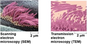
Scanning Electron Microscope (SEM): Provides 3D images of specimen surfaces.
Transmission Electron Microscope (TEM): Used to study internal cell structures by passing electrons through thin sections of specimens.
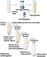
Cell Fractionation
Cell fractionation is a technique used to separate cellular components for individual study. It involves homogenizing cells and using centrifugation at various speeds to isolate organelles based on size and density.
Allows correlation of cell structure with function.
Enables biochemical analysis of organelles.
Cell Types: Prokaryotic vs. Eukaryotic
Basic Features of All Cells
All cells share certain features:
Plasma membrane: Selective barrier for passage of materials.
Cytosol: Semifluid substance where subcellular components are suspended.
Chromosomes: Carry genetic information.
Ribosomes: Synthesize proteins.
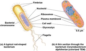
Prokaryotic Cells
Prokaryotic cells (Bacteria and Archaea) lack a nucleus and membrane-bound organelles. Their DNA is located in the nucleoid region, and the cytoplasm is bound by the plasma membrane.
Eukaryotic Cells
Eukaryotic cells (protists, fungi, animals, plants) have a nucleus enclosed by a double membrane and various membrane-bound organelles. They are generally larger than prokaryotic cells.
Surface Area to Volume Ratio
The ratio of surface area to volume is critical for cell function. As cells increase in size, their volume grows faster than their surface area, limiting the efficiency of material exchange.
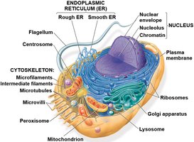
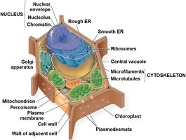
Internal Organization of Eukaryotic Cells
Nucleus and Ribosomes
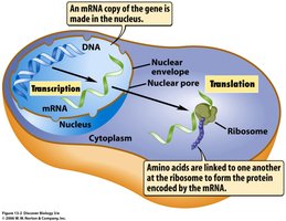
The nucleus contains most of the cell's genetic material and is surrounded by the nuclear envelope, a double membrane with pores for molecular exchange. The nucleolus within the nucleus is the site of ribosomal RNA synthesis and ribosome assembly.
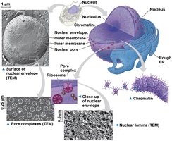
DNA is organized into chromosomes, which are made of chromatin (DNA and proteins). The process of gene expression involves transcription (DNA to mRNA) in the nucleus and translation (mRNA to protein) in the cytoplasm at ribosomes.
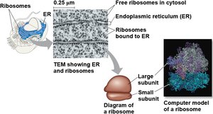
Ribosomes
Ribosomes are complexes of rRNA and protein that synthesize proteins. They can be free in the cytosol or bound to the endoplasmic reticulum (ER) or nuclear envelope.
Free ribosomes produce proteins for use within the cytosol.
Bound ribosomes produce proteins for membranes, organelles, or export.
The Endomembrane System
Components and Functions
The endomembrane system includes the nuclear envelope, ER, Golgi apparatus, lysosomes, vacuoles, and plasma membrane. It is involved in protein synthesis, transport, metabolism, and detoxification.
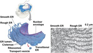
Endoplasmic Reticulum (ER)
The ER is a network of membranes with two regions:
Smooth ER: Lacks ribosomes; synthesizes lipids, detoxifies drugs, stores calcium ions.
Rough ER: Studded with ribosomes; modifies and packages proteins, produces membranes.
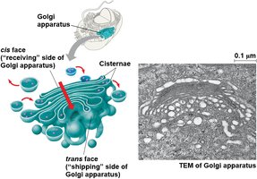
Golgi Apparatus
The Golgi apparatus consists of flattened sacs (cisternae) and functions as the cell's shipping and receiving center. It modifies, sorts, and packages proteins and lipids for delivery to various destinations.
Cis face: Receiving side
Trans face: Shipping side

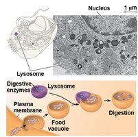
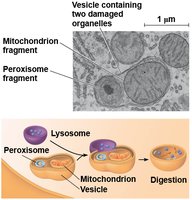
Lysosomes
Lysosomes are membrane-bound sacs containing hydrolytic enzymes for digesting macromolecules. They are involved in phagocytosis (ingestion of material) and autophagy (recycling of cellular components).
Defects in lysosomal enzymes can cause storage diseases (e.g., Tay-Sachs disease).
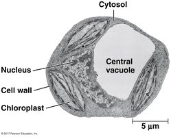
Vacuoles
Vacuoles are large vesicles with diverse functions, including storage, waste disposal, and maintaining cell turgor. Central vacuoles in plant cells store water and ions and play a major role in cell growth.
Energy Conversion Organelles
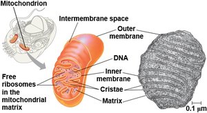
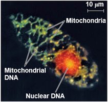
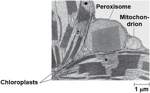
Mitochondria
Mitochondria are the sites of cellular respiration, converting energy from organic molecules into ATP. They have a double membrane, their own DNA, and ribosomes, supporting the endosymbiont theory of their origin.
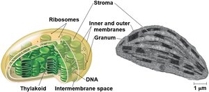
Chloroplasts
Chloroplasts are found in plants and algae and are the sites of photosynthesis. They contain chlorophyll, thylakoids (stacked into grana), and stroma, and have their own DNA and ribosomes.
Peroxisomes
Peroxisomes are single-membrane organelles that carry out oxidation reactions, producing hydrogen peroxide and converting it to water. They are involved in fatty acid breakdown and detoxification.
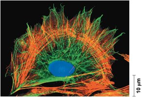
The Cytoskeleton
Structure and Function
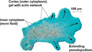
The cytoskeleton is a network of protein fibers that provides structural support, organizes cell components, and enables cell movement. It consists of microtubules, microfilaments, and intermediate filaments.
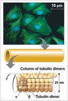
Microtubules
Microtubules are hollow rods made of tubulin dimers. They maintain cell shape, guide organelle movement, and are involved in chromosome separation during cell division and in the structure of cilia and flagella.
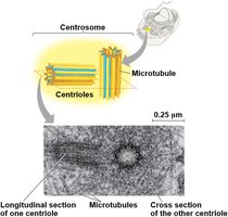
Centrosomes and Centrioles
In animal cells, microtubules grow from the centrosome, which contains a pair of centrioles. These structures help organize microtubule assembly.
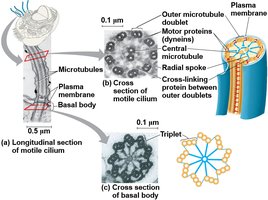
Cilia and Flagella
Cilia and flagella are microtubule-containing extensions that enable cell movement. They have a characteristic "9+2" arrangement of microtubules and are anchored by a basal body. The motor protein dynein drives their movement.

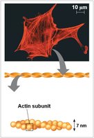
Microfilaments (Actin Filaments)
Microfilaments are thin rods of actin that support cell shape, enable cell movement (e.g., muscle contraction, amoeboid movement), and drive cytoplasmic streaming in plant cells.
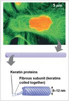
Intermediate Filaments
Intermediate filaments are fibrous proteins (e.g., keratin) that provide mechanical support and help anchor organelles. They are more permanent than microtubules or microfilaments.
Extracellular Structures and Cell Junctions
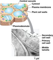
Cell Walls of Plants
Plant cell walls are composed of cellulose and provide structural support, protection, and regulation of water uptake. They may have multiple layers: primary cell wall, middle lamella, and secondary cell wall.
Extracellular Matrix (ECM) of Animal Cells
The ECM is a network of glycoproteins (collagen, proteoglycans, fibronectin) that supports animal cells, helps bind them together, and transmits signals via integrins to the cytoskeleton.
Cell Junctions
Cells in tissues are connected by specialized junctions:
Tight junctions: Seal cells together to prevent leakage.
Desmosomes: Anchor cells together into strong sheets.
Gap junctions: Allow communication by permitting the passage of small molecules between cells.
Plasmodesmata: Channels in plant cell walls that connect the cytoplasm of adjacent cells.
Integration of Cellular Structures
Cellular function arises from the integration and coordination of all cellular structures and organelles. No compartment works alone; for example, processes like phagocytosis require the cytoskeleton, lysosomes, and plasma membrane to work together.
