 Back
BackA Tour of the Cell: Structure, Function, and Diversity
Study Guide - Smart Notes
 Tailored notes based on your materials, expanded with key definitions, examples, and context.
Chapter 6: A Tour of the Cell
Introduction to Cell Diversity
Cells are the fundamental units of life, exhibiting remarkable diversity in their structures, macromolecules, and processes. This diversity underpins the complexity and specialization seen in living organisms.
History of Cell Discovery
Early Microscopy and Cell Theory
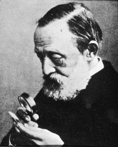
Robert Hooke (1665): First to use a simple microscope to observe cells, coining the term "cellulae."
Antonie van Leeuwenhoek (1680s): Improved microscopes and observed microorganisms in water.
Microscopy revealed the presence and detailed structure of cells.
Magnification: Ratio of image size to actual size.
Resolution: Measure of image clarity.
Contrast: Difference in brightness between light and dark areas.
Microscopy Techniques
Types of Microscopes
Light Microscopes: Magnify up to 1000x, suitable for viewing live cells.
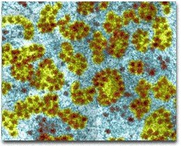
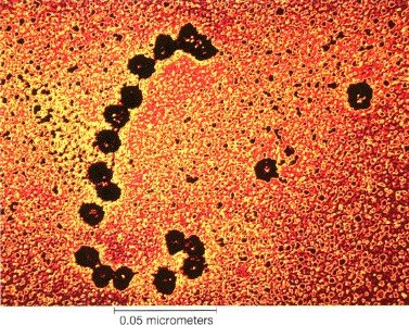
Transmission Electron Microscopes (TEM): Use electrons to visualize internal cell structures, up to 500,000x magnification.
Scanning Electron Microscopes (SEM): Provide 3D images of cell surfaces.
Cryo-electron Microscopy: Preserves specimens in a near-native state for imaging.
Cell Fractionation: Technique to separate cellular components for study.
Prokaryotic vs. Eukaryotic Cells
Key Differences
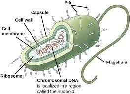
Prokaryotic Cells: Lack a nucleus; DNA is in a nucleoid region. No membrane-bound organelles. Examples: Bacteria and Archaea.
Eukaryotic Cells: Have a true nucleus and many membrane-bound organelles. Found in animals, plants, fungi, and protists.
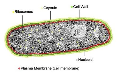
Prokaryotic Cell Structure
Nucleoid: Region containing DNA, not membrane-bound.
Cell Wall: Provides structure and protection.
Capsule/Slime Layer: Offers additional protection.
Ribosomes: Sites of protein synthesis.
Lack of membrane-bound organelles.
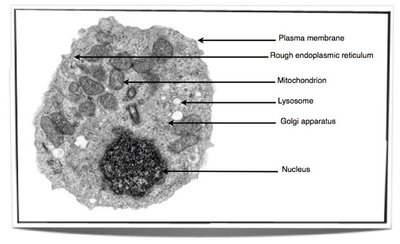
Eukaryotic Cell Structure
Nucleus: Contains chromosomes, separated from cytoplasm by nuclear envelope.
Membrane-bound Organelles: Specialized compartments for various functions.
Present in animals, plants, fungi, and protists.
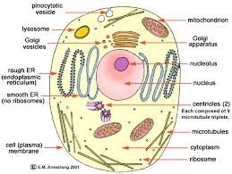
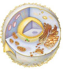
Cytoplasm and Organelles
Cytoplasm
Jelly-like interior of the cell where organelles are suspended.
Cytosol: Watery component of the cytoplasm.
Organelles
Specialized structures with specific functions (e.g., mitochondria, Golgi apparatus, lysosomes).
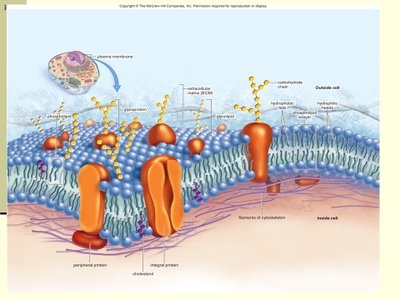
Plasma Membrane
Structure and Function
Phospholipid bilayer with embedded proteins.
Acts as a selective barrier, controlling entry and exit of substances.
Separates cell contents from the environment.
Nucleus and Genetic Material
Nucleus
Enclosed by a double membrane (nuclear envelope) with pores.
Contains chromosomes and nucleoli.
Controls cellular reproduction and protein synthesis.
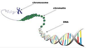
Chromosomes and Chromatin
Chromatin: DNA-protein complex, appears grainy except during cell division.
Condenses into chromosomes before cell division.
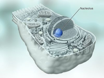
Nucleolus
Dense region within the nucleus.
Site of ribosome synthesis (rRNA and proteins).
Cells may have more than one nucleolus.
Ribosomes
Structure and Function
Composed of rRNA and proteins.
Sites of protein synthesis.
Can be free in cytosol or bound to endoplasmic reticulum.
Polyribosome: Group of ribosomes translating the same mRNA.
Endomembrane System
Components and Functions
Includes nuclear envelope, endoplasmic reticulum (ER), Golgi apparatus, lysosomes, vesicles, vacuoles, and plasma membrane.
Membranes may be continuous or connected via vesicles.
Absent in prokaryotic cells.
Endoplasmic Reticulum (ER)
Network of membranous channels and sacs connected to the nuclear envelope.
Rough ER: Studded with ribosomes; synthesizes proteins for export and membrane.
Smooth ER: Lacks ribosomes; synthesizes lipids, detoxifies drugs, stores Ca2+, and metabolizes carbohydrates.
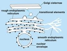
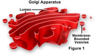
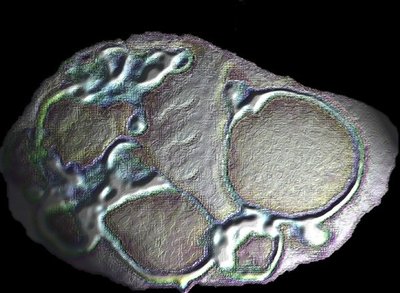
Golgi Apparatus
Stack of flattened membranous sacs (cisternae).
Processes, packages, and secretes proteins and lipids.
Cis face receives, trans face ships out vesicles.
Synthesizes some polysaccharides.
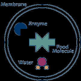
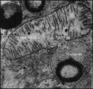
Lysosomes
Membranous vesicles containing hydrolytic enzymes.
Function in intracellular digestion (autophagy) and defense against pathogens.
Sometimes called "suicide bags" due to their role in cell death.
Vacuoles
Membrane-bound sacs for storage and transport.
Food vacuoles, contractile vacuoles (in protists), and central vacuole (in plants).
Central vacuole stores organic compounds and pigments, and supports plant cell growth.
Energy-Related Organelles
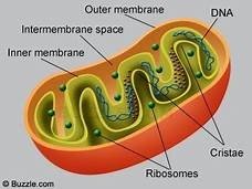
Mitochondria
Site of aerobic cellular respiration:
Double membrane; inner membrane forms cristae, matrix is the inner fluid space.
Contain their own DNA and ribosomes.
Known as the "powerhouse of the cell."
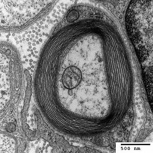
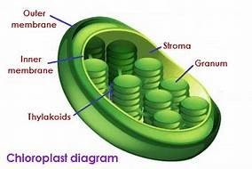
Chloroplasts
Site of photosynthesis:
Contain chlorophyll, thylakoids (stacked into grana), and stroma (fluid-filled space).
Other plastids include amyloplasts (starch storage) and chromoplasts (pigment storage).
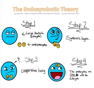
Endosymbiotic Theory
Explains the origin of mitochondria and chloroplasts as descendants of engulfed prokaryotes.
Both organelles contain their own DNA and ribosomes, supporting this theory.
Other Organelles
Peroxisomes
Single-membrane organelles containing enzymes that transfer hydrogen to oxygen, forming hydrogen peroxide (), which is then converted to water.
Break down fatty acids and detoxify alcohol in liver cells.
Cytoskeleton
Structure and Function
Network of protein fibers that provide cell shape, support, and movement.
Composed of microtubules (thick), intermediate filaments (medium), and microfilaments (thin).
Microtubules
Hollow rods made of tubulin.
Maintain cell shape, facilitate movement of organelles and vesicles, and form the spindle apparatus during mitosis.
Make up centrioles, cilia, and flagella (9+2 arrangement in cilia/flagella).
Microfilaments and Intermediate Filaments
Microfilaments: Thin rods of actin; involved in muscle contraction, cell movement, and cytoplasmic streaming.
Intermediate Filaments: Provide structural support; anchor organelles and maintain cell integrity.
Cell Wall and Cell Junctions
Cell Wall
Present in plants, fungi, and some protists.
Composed mainly of cellulose in plants; provides protection and structural support.
Cell Junctions
Plasmodesmata: Channels connecting plant cells, allowing transport and communication.
Tight Junctions, Desmosomes, Gap Junctions: Specialized connections in animal cells, especially common in epithelial tissue.
