 Back
BackA Tour of the Cell: Structure, Function, and Diversity
Study Guide - Smart Notes
 Tailored notes based on your materials, expanded with key definitions, examples, and context.
A Tour of the Cell
Introduction to Cell Structure
Cells are the fundamental units of life, and all living organisms are composed of cells. Each cell contains essential biomolecules and structures that enable it to function, grow, and reproduce. The main components found in all cells include nucleic acids, proteins, carbohydrates, and a plasma membrane.
Nucleic acids: Store and transmit genetic information (DNA and RNA).
Proteins: Perform most cellular functions, including catalysis, structure, and signaling.
Carbohydrates: Provide energy, structural support, and cellular identity.
Plasma membrane: Acts as a selectively permeable barrier, regulating the entry and exit of substances.
Classification of Cells
Cells are classified based on their morphology and evolutionary history. Morphologically, cells are grouped as prokaryotes or eukaryotes. Phylogenetically, life is divided into three domains: Bacteria, Archaea, and Eukarya.
Prokaryotes: Lack a membrane-bound nucleus; include Bacteria and Archaea.
Eukaryotes: Possess a membrane-bound nucleus; include Eukarya.
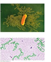
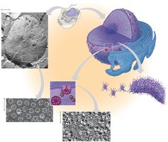
Prokaryotic Cells
Structure and Components
Prokaryotic cells are generally smaller and simpler than eukaryotic cells. They lack membrane-bound organelles but possess specialized structures for survival and function.
Nucleoid: Region containing the cell's DNA, not enclosed by a membrane.
Ribosomes: Sites of protein synthesis.
Plasma membrane: Encloses the cytoplasm.
Cell wall: Provides rigidity and protection; composed of peptidoglycan in bacteria.
Glycocalyx: Outer coating, either a capsule or slime layer, for protection and adhesion.
Fimbriae: Attachment structures on the surface.
Flagella: Locomotion organelles.
Example: Corynebacterium diphtheriae is a rod-shaped bacterium with a visible nucleoid and cell wall.
Genetic Material in Prokaryotes
Prokaryotic DNA is typically a single, circular chromosome located in the nucleoid. DNA is often supercoiled to fit within the cell. Plasmids are small, circular DNA molecules that carry additional genes.
Eukaryotic Cells
Key Differences from Prokaryotes
Eukaryotic cells are larger and more complex, with extensive internal membranes and organelles. Four key differences include:
Chromosomes are enclosed within a membrane-bound nucleus.
Cells are often much larger.
Contain numerous organelles and internal membranes, increasing efficiency and compartmentalization.
Feature a dynamic cytoskeleton for structural support and movement.
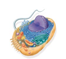
Animal Cell Structure
Animal cells contain a variety of organelles, each with specialized functions:
Nucleus: Contains chromatin, nucleolus, and nuclear envelope.
Endoplasmic Reticulum (ER): Rough ER (protein synthesis), Smooth ER (lipid synthesis, Ca2+ storage).
Golgi apparatus: Modifies, sorts, and packages proteins and lipids.
Mitochondrion: Site of cellular respiration and ATP production.
Lysosome: Digests macromolecules and damaged organelles.
Peroxisome: Metabolizes hydrogen peroxide.
Centrosome: Initiates microtubules.
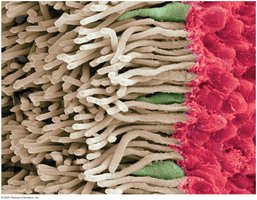
Microvilli: Increase surface area.
Cytoskeleton: Provides structural support and facilitates movement.
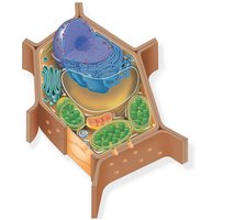
Plant Cell Structure
Plant cells share many organelles with animal cells but also possess unique structures:
Cell wall: Provides structural support and protection.
Chloroplast: Site of photosynthesis.
Central vacuole: Storage, waste breakdown, and hydrolysis.
Plasmodesmata: Channels connecting adjacent cells.
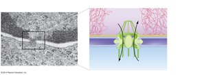
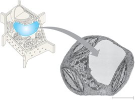
The Nucleus and Its Envelope
Structure and Function
The nucleus is the control center of the cell, housing genetic material and coordinating activities such as growth and reproduction. The nuclear envelope is a double membrane with nuclear pores for molecular transport.
Chromatin: DNA-protein complex, exists in varying states of coiling.
Nucleolus: Site of ribosome production.
Nuclear lamina: Netlike structure supporting the envelope.
Nuclear pores: Allow selective import/export of molecules.
Nuclear Import
Proteins destined for the nucleus contain a nuclear localization signal (NLS), a specific amino acid sequence that acts as a molecular zip code for import.
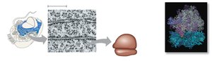
Ribosomes
Structure and Function
Ribosomes are the molecular machines responsible for protein synthesis. They can be free in the cytosol or bound to the ER.
Composed of a large and small subunit.
Translate mRNA into polypeptides.
Endoplasmic Reticulum (ER)
Rough and Smooth ER
The ER is a network of membranous sacs and tubes. The rough ER is studded with ribosomes and synthesizes proteins, while the smooth ER is involved in lipid synthesis and Ca2+ storage.
Rough ER: Protein synthesis and folding.
Smooth ER: Lipid metabolism, detoxification, and Ca2+ reservoir.
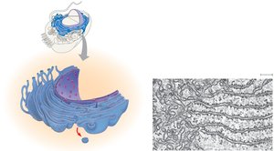
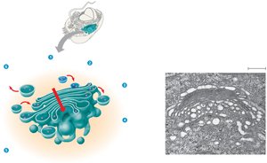
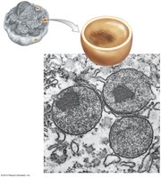
Golgi Apparatus
Structure and Function
The Golgi apparatus is a series of flattened membranous sacs (cisternae) that modify, sort, and package proteins and lipids for secretion or delivery to other organelles.
Has a cis face (receiving side) and trans face (shipping side).
Proteins move through the Golgi via vesicles and cisternal maturation.
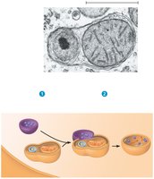
Lysosomes
Autophagy and Phagocytosis
Lysosomes are digestive organelles containing hydrolytic enzymes. They break down macromolecules, damaged organelles (autophagy), and engulfed particles (phagocytosis).
Fuse with vesicles containing material to be digested.
Recycle cellular components.
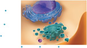
Endomembrane System
Relationships Among Organelles
The endomembrane system includes the nuclear envelope, ER, Golgi apparatus, lysosomes, and vesicles. It coordinates the synthesis, modification, and transport of proteins and lipids.
Membranes and proteins produced by the ER move via vesicles to the Golgi.
The Golgi sorts and packages proteins for secretion or delivery to other organelles.
Lysosomes and vacuoles arise from the Golgi.
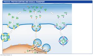
Protein Sorting and Secretion
Proteins are tagged, sorted, and delivered to their destinations via vesicle transport. The secretory pathway involves the ER, Golgi, and plasma membrane.
Proteins are tagged for sorting.
Vesicles bud and deliver proteins to the plasma membrane or other organelles.
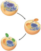
Plant Cell Vacuole
Structure and Function
The central vacuole in plant cells is a large organelle responsible for storage, waste breakdown, and hydrolysis. It also contributes to cell growth by enlarging.
Peroxisomes
Structure and Function
Peroxisomes are organelles with specialized metabolic functions, including the breakdown of fatty acids and detoxification of hydrogen peroxide.
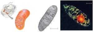
Mitochondria
Site of Cellular Respiration
Mitochondria are double-membraned organelles where cellular respiration occurs, generating ATP. They contain their own DNA and ribosomes.
Inner membrane: Highly folded (cristae) for increased surface area.
Matrix: Contains enzymes, DNA, and ribosomes.
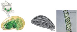
Chloroplasts
Site of Photosynthesis
Chloroplasts are found in plant cells and are responsible for photosynthesis. They contain thylakoids, stroma, and their own DNA and ribosomes.
Thylakoids: Membranous sacs where light reactions occur.
Stroma: Fluid surrounding thylakoids, site of Calvin cycle.
Endosymbiont Theory
Origin of Mitochondria and Chloroplasts
The endosymbiont theory proposes that mitochondria and chloroplasts originated from prokaryotic cells engulfed by ancestral eukaryotes. Evidence includes their double membranes, DNA, and ribosomes.
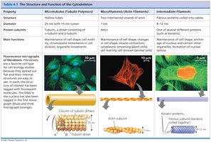
Cytoskeleton
Structure and Function
The cytoskeleton is a network of protein filaments that provides structural support, facilitates movement, and organizes cellular components. It consists of microfilaments, intermediate filaments, and microtubules.
Microfilaments (Actin filaments): Define cell shape, involved in muscle movement, cytokinesis, and cytoplasmic streaming.
Intermediate filaments: Provide mechanical support and maintain cell shape.
Microtubules: Hollow tubes involved in cell shape, movement, and organelle transport.
Motor Proteins
Motor proteins interact with the cytoskeleton to transport vesicles and organelles within the cell. They use ATP to "walk" along microtubules or microfilaments.
Flagella and Cilia
Flagella and cilia are motile structures composed of microtubules arranged in a "9+2" pattern. They facilitate cell movement and are powered by motor proteins (dyneins).
Cell Wall and Extracellular Matrix
Cell Wall
Plants, algae, and fungi possess a cell wall that provides structural support and protection. The composition varies among groups:
Plants and algae: Cellulose and polysaccharides.
Fungi: Chitin.
Bacteria: Peptidoglycan.
Extracellular Matrix (ECM) in Animal Cells
The ECM is a network of proteins and carbohydrates outside animal cells, providing structural support and facilitating cell signaling.
Collagen: Main structural protein.
Proteoglycans: Protein-polysaccharide complexes.
Fibronectin: Attaches ECM to integrins in the plasma membrane.
Integrins: Transmembrane proteins linking ECM to cytoskeleton.
Cell Junctions
Animal Cell Junctions
Cell junctions facilitate communication and adhesion between animal cells:
Tight junctions: Prevent fluid movement between cells.
Desmosomes: Provide mechanical strength.
Gap junctions: Allow passage of ions and small molecules.
Plasmodesmata in Plant Cells
Plasmodesmata are channels that connect plant cells, allowing transport of molecules and communication.
Dynamic Nature of Eukaryotic Cells
Cellular Activity and Renewal
Eukaryotic cells are highly dynamic, constantly synthesizing ATP, catalyzing reactions, and renewing organelles such as mitochondria. Membrane composition is continually changing to adapt to cellular needs.
Cells synthesize ~10 million ATP molecules per second.
Enzymes catalyze >25,000 reactions per second.
Mitochondria are replaced every 10 days.
