 Back
BackA Tour of the Cell: Structure, Function, and Microscopy
Study Guide - Smart Notes
 Tailored notes based on your materials, expanded with key definitions, examples, and context.
The Cell: An Overview
Introduction to Cell Biology
Cells are the fundamental units of life, forming the basis of all living organisms. The study of cells, their structure, and their function is central to understanding biology. Cells can be visualized using various types of microscopy, each providing unique insights into cellular architecture and processes.
Microscopy and Cell Visualization
Types of Microscopy
Light Microscopy (LM): Uses visible light to illuminate specimens, allowing observation of living cells and tissues.
Electron Microscopy (EM): Employs electron beams for much higher resolution, revealing ultrastructural details of cells.
Super-Resolution Microscopy: Advanced techniques that surpass the resolution limits of conventional light microscopy.
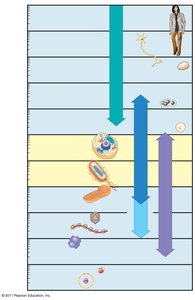
Microscopy enables the study of cell size, structure, and organization, from atoms and small molecules to entire cells and tissues.
Microscopy Techniques and Applications
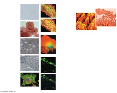
Brightfield Microscopy: Standard light microscopy, can be used with or without stains.
Phase-Contrast and Differential-Interference-Contrast: Enhance contrast in unstained cells.
Fluorescence Microscopy: Uses fluorescent dyes or proteins to label specific cell components.
Confocal and Deconvolution Microscopy: Provide optical sectioning for detailed 3D reconstructions.
Scanning Electron Microscopy (SEM): Visualizes cell surfaces in high detail.
Transmission Electron Microscopy (TEM): Reveals internal cell structures at high resolution.
Types of Cells: Prokaryotic and Eukaryotic
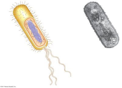
Prokaryotic Cells
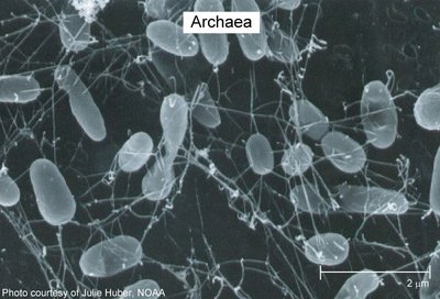
Prokaryotic cells are simpler and smaller than eukaryotic cells. They lack a nucleus and membrane-bound organelles. Their DNA is located in a region called the nucleoid. Prokaryotes include organisms from the domains Bacteria and Archaea.
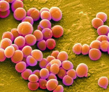
Key Features: Plasma membrane, cytosol, chromosomes, ribosomes, cell wall, sometimes capsule, fimbriae, and flagella.
Example: Staphylococcus aureus (bacterium), Archaea.
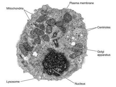
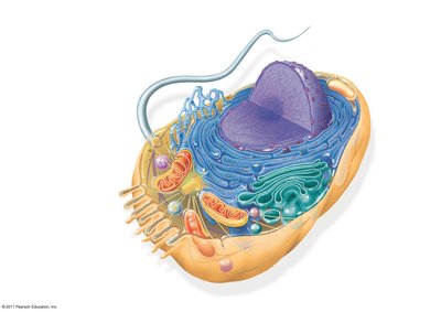
Eukaryotic Cells
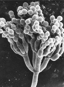
Eukaryotic cells are more complex, with DNA enclosed in a nucleus and numerous membrane-bound organelles. They are found in protists, fungi, animals, and plants.
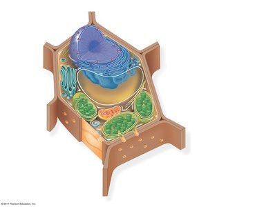
Key Features: Nucleus, endoplasmic reticulum, Golgi apparatus, mitochondria, lysosomes, peroxisomes, cytoskeleton, and in plants, chloroplasts and a central vacuole.
Examples: Animal cells, plant cells, fungi (e.g., Penicillium), protists.
Cell Structure and Function
Basic Features of All Cells
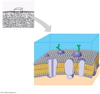
Plasma Membrane: Selective barrier composed of a phospholipid bilayer with embedded proteins, controlling the movement of substances in and out of the cell.
Cytosol: Semifluid substance within the cell where organelles are suspended.
Chromosomes: Carry genetic information (DNA).
Ribosomes: Sites of protein synthesis.
Plasma Membrane Structure
The plasma membrane is a dynamic structure that separates the cell from its environment and regulates the passage of materials. It consists of a double layer of phospholipids with embedded proteins and carbohydrate side chains.
Internal Organization of Eukaryotic Cells
Animal and Plant Cell Structures
Eukaryotic cells contain a variety of organelles, each with specialized functions. Plant and animal cells share many organelles, but plant cells also have a cell wall, chloroplasts, and a large central vacuole.
The Nucleus
The nucleus houses most of the cell’s genetic material. It is surrounded by a double membrane called the nuclear envelope, which contains nuclear pores for molecular transport. The nucleolus within the nucleus is the site of ribosomal RNA (rRNA) synthesis.
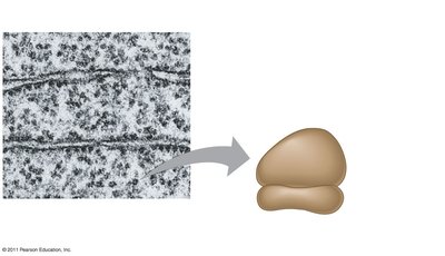
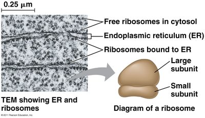
Ribosomes: Protein Factories
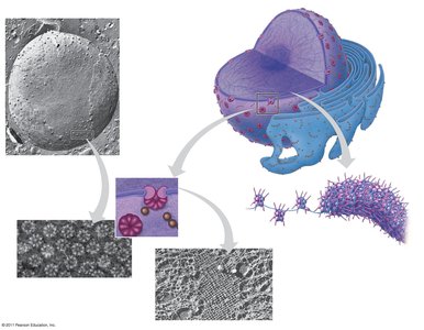
Ribosomes are complexes of rRNA and protein that synthesize proteins. They can be free in the cytosol or bound to the endoplasmic reticulum (ER) or nuclear envelope.
The Endomembrane System
The endomembrane system is a network of membranes within eukaryotic cells that regulates protein traffic and performs metabolic functions. It includes the nuclear envelope, ER, Golgi apparatus, lysosomes, vacuoles, and plasma membrane.
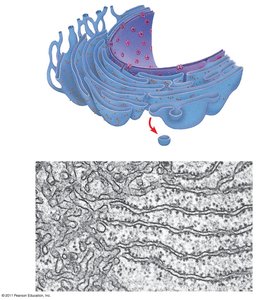
Endoplasmic Reticulum (ER)
Smooth ER: Lacks ribosomes; synthesizes lipids, metabolizes carbohydrates, detoxifies drugs, and stores calcium ions.
Rough ER: Studded with ribosomes; synthesizes proteins and membranes, distributes transport vesicles.
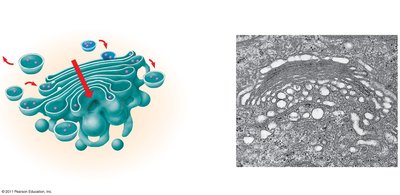
Golgi Apparatus
The Golgi apparatus modifies, sorts, and packages proteins and lipids for secretion or delivery to other organelles. It has a cis face (receiving side) and a trans face (shipping side).
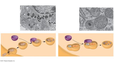
Lysosomes
Lysosomes are membrane-bound sacs containing hydrolytic enzymes for digesting macromolecules. They are involved in phagocytosis (ingestion of external particles) and autophagy (recycling of cellular components).
Vacuoles
Food Vacuoles: Formed by phagocytosis in some cells.
Contractile Vacuoles: Pump excess water out of cells (common in protists).
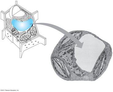
Central Vacuole: Found in plant cells, stores water and organic compounds.
Energy-Transforming Organelles
Mitochondria
Mitochondria are the sites of cellular respiration, generating ATP from organic molecules. They have a double membrane, with the inner membrane folded into cristae to increase surface area for ATP synthesis. Mitochondria contain their own DNA and ribosomes.
Chloroplasts
Chloroplasts are found in plants and algae and are the sites of photosynthesis. They contain the pigment chlorophyll, thylakoid membranes (stacked into grana), and stroma (internal fluid). Chloroplasts also have their own DNA and ribosomes.
Peroxisomes
Peroxisomes are specialized organelles that carry out oxidation reactions, producing hydrogen peroxide and converting it to water. They are involved in lipid metabolism and detoxification.
The Cytoskeleton
Structure and Function
The cytoskeleton is a network of protein fibers that provides structural support, maintains cell shape, and facilitates cell movement and intracellular transport. It interacts with motor proteins to move vesicles and organelles within the cell.
Types of Cytoskeletal Fibers
Microtubules: Hollow rods made of tubulin; involved in cell shape, organelle movement, and chromosome separation during cell division.
Microfilaments (Actin Filaments): Solid rods of actin; support cell shape, involved in muscle contraction and cell movement.
Intermediate Filaments: Fibrous proteins; provide mechanical support and anchor organelles.
Extracellular Components and Cell Junctions
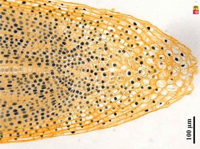
Cell Walls of Plants
Plant cell walls are composed of cellulose and provide structural support, protection, and regulation of water uptake. They are distinct from the plasma membrane and are found in prokaryotes, fungi, and some protists as well.
Extracellular Matrix (ECM) of Animal Cells
The ECM is a network of glycoproteins (such as collagen, proteoglycans, and fibronectin) that provides structural and biochemical support to animal cells. ECM proteins interact with cell surface receptors called integrins, influencing cell behavior and tissue organization.
Cell Junctions
Plasmodesmata: Channels in plant cell walls that allow transport of water, ions, and small molecules between cells.
Tight Junctions: Seal adjacent animal cells to prevent leakage of extracellular fluid.
Desmosomes: Anchor animal cells together into strong sheets.
Gap Junctions: Provide cytoplasmic channels for communication between adjacent animal cells.
Summary Table: Prokaryotic vs. Eukaryotic Cells
Feature | Prokaryotic Cells | Eukaryotic Cells |
|---|---|---|
Nucleus | Absent | Present |
Membrane-bound Organelles | Absent | Present |
Size | 1–10 μm | 10–100 μm |
Domains | Bacteria, Archaea | Eukarya (Protists, Fungi, Plants, Animals) |
Examples | Staphylococcus, Archaea | Animal, plant, fungal, protist cells |
Additional info: This guide covers the main concepts from "A Tour of the Cell" (Ch. 6), including cell structure, microscopy, and the differences between prokaryotic and eukaryotic cells, as well as the organization and function of cellular organelles and extracellular components.
