 Back
A Tour of the Cell: Structure and Function of Cellular Components
Study Guide - Smart Notes
 Tailored notes based on your materials, expanded with key definitions, examples, and context.
Introduction to the Cell
Historical Perspective and Cell Theory
The development of microscopy revolutionized our understanding of cells, the fundamental units of life. Early observations by Robert Hooke and Antonie van Leeuwenhoek laid the foundation for modern cell biology. The cell theory, a cornerstone of biology, states that all living things are composed of cells and all cells arise from pre-existing cells.
Cell: The smallest unit of life, capable of carrying out all life processes.
Cell Theory: All organisms are made of cells; all cells come from other cells.
Microscopy: Essential for visualizing cells and their structures.
Microscopes Reveal the World of the Cell
Types of Microscopes and Their Applications
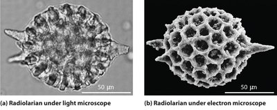
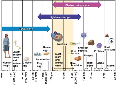
Microscopes are indispensable tools for studying cells. The light microscope allows observation of living cells, while scanning electron microscopes (SEM) and transmission electron microscopes (TEM) provide much greater magnification and resolution, revealing fine details of cell structure.
Magnification: The increase in an object's image size compared to its actual size.
Resolution: The clarity of an image; the ability to distinguish two close objects as separate.
Light Microscope (LM): Suitable for viewing living cells and general cell structure.
Scanning Electron Microscope (SEM): Used for detailed surface structures.
Transmission Electron Microscope (TEM): Used for viewing internal cell structures at high resolution.
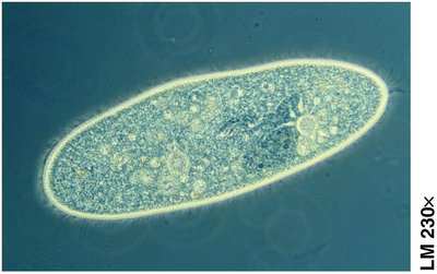
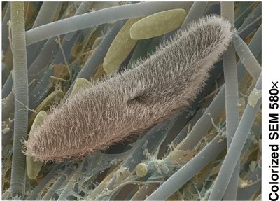
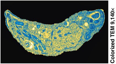
Example: To observe the shape changes of a living white blood cell, use a light microscope. For surface texture of a hair, use SEM. For organelle details, use TEM.
The Small Size of Cells and the Plasma Membrane
Surface-to-Volume Ratio and Membrane Structure
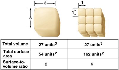
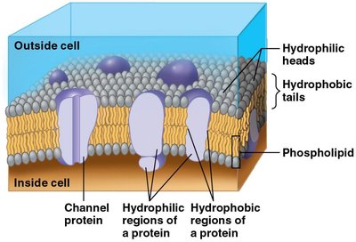
Cells are small to maximize their surface-to-volume ratio, facilitating efficient exchange of materials with their environment. The plasma membrane is a phospholipid bilayer with embedded proteins, crucial for regulating the movement of substances in and out of the cell.
Surface-to-Volume Ratio: Smaller cells have a higher ratio, allowing more efficient exchange.
Plasma Membrane: Composed of a double layer of phospholipids with proteins that act as channels or pumps.
Prokaryotic and Eukaryotic Cells
Structural Differences and Domains of Life
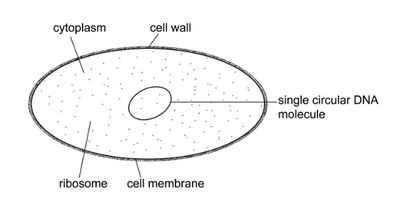
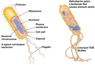
Cells are classified as prokaryotic or eukaryotic. Prokaryotic cells (Bacteria and Archaea) lack a nucleus and membrane-bound organelles, while eukaryotic cells (Eukarya) possess these structures.
Prokaryotic Cells: Simpler, smaller, no nucleus, DNA in nucleoid region.
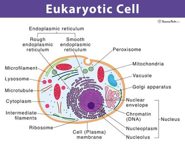
Eukaryotic Cells: Larger, complex, nucleus, and various organelles.
Functional Compartments in Eukaryotic Cells
Organization of Cellular Functions
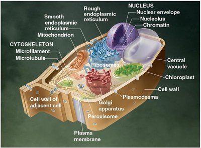
Eukaryotic cells are divided into compartments, each specialized for distinct functions. These include genetic control, manufacturing and breakdown, energy processing, and structural support/communication.
Genetic Control: Nucleus and ribosomes.
Manufacturing, Distribution, Breakdown: Endoplasmic reticulum, Golgi apparatus, lysosomes, vacuoles.
Energy Processing: Mitochondria (all cells), chloroplasts (plants/algae).
Structural Support, Movement, Communication: Cytoskeleton, plasma membrane, cell wall (plants).
The Nucleus and Ribosomes
Genetic Information and Protein Synthesis
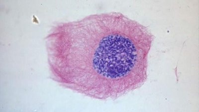
The nucleus contains the cell's DNA and directs protein synthesis via messenger RNA (mRNA). Ribosomes, assembled in the nucleolus, are the sites of protein synthesis.
Nucleus: Site of DNA replication, transcription, and ribosome assembly.
Ribosomes: Composed of rRNA and proteins; synthesize proteins according to genetic instructions.
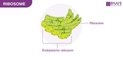
Free vs. Bound Ribosomes: Free ribosomes function in the cytosol; bound ribosomes are attached to the endoplasmic reticulum.
The Endomembrane System
Membrane-Bound Organelles and Their Functions
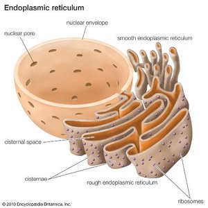
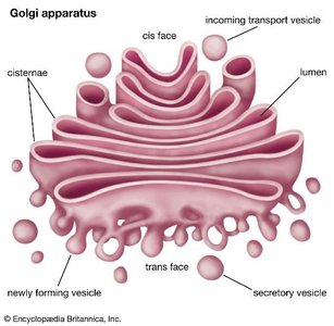
The endomembrane system includes the nuclear envelope, endoplasmic reticulum (ER), Golgi apparatus, lysosomes, vacuoles, and plasma membrane. These organelles work together in the synthesis, modification, packaging, and transport of cellular materials.
Endoplasmic Reticulum (ER): Rough ER (with ribosomes) synthesizes proteins; smooth ER synthesizes lipids and detoxifies chemicals.
Golgi Apparatus: Modifies, sorts, and ships proteins and lipids received from the ER.
Lysosomes: Contain digestive enzymes to break down macromolecules and damaged organelles.
Vacuoles: Large vesicles for storage and maintenance; central vacuole in plants regulates water and stores substances.
Peroxisomes: Metabolic compartments not derived from the endomembrane system; break down fatty acids and detoxify harmful substances.
Energy-Converting Organelles
Mitochondria and Chloroplasts
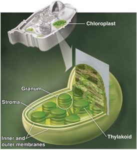
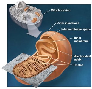
Mitochondria are the sites of cellular respiration, converting energy from food into ATP. Chloroplasts (in plants and algae) carry out photosynthesis, converting solar energy into chemical energy stored in sugars.
Mitochondria: Have an outer membrane, inner membrane (with cristae), intermembrane space, and matrix.
Chloroplasts: Contain inner and outer membranes, stroma, thylakoids, and grana.
Endosymbiosis Theory: Mitochondria and chloroplasts originated as free-living prokaryotes engulfed by ancestral eukaryotic cells.
Contrasting Plant and Animal Cells
Unique Features of Plant and Animal Cells
Plant and animal cells share many organelles but also have distinct structures. Plant cells possess a cell wall, chloroplasts, and a large central vacuole, while animal cells have centrosomes and lysosomes.
Plant Cells: Cell wall (mainly cellulose), chloroplasts, central vacuole.
Animal Cells: Centrosome (with centrioles), lysosomes.
Summary Table: Eukaryotic Cell Structures and Their Functions
Structure | Main Function |
|---|---|
Nucleus | Houses genetic material, controls cell activities |
Ribosome | Protein synthesis |
Endoplasmic Reticulum (Rough/Smooth) | Protein and lipid synthesis, detoxification |
Golgi Apparatus | Modification, sorting, and shipping of proteins/lipids |
Lysosome | Digestion of macromolecules and old organelles |
Vacuole | Storage, water regulation, waste disposal |
Mitochondrion | ATP production via cellular respiration |
Chloroplast | Photosynthesis (plants/algae) |
Peroxisome | Breakdown of fatty acids, detoxification |
Cytoskeleton | Structural support, movement |
Plasma Membrane | Regulates entry/exit of substances |
Cell Wall (plants) | Protection, structural support |
