 Back
BackA Tour of the Cell: Structure, Function, and Diversity
Study Guide - Smart Notes
 Tailored notes based on your materials, expanded with key definitions, examples, and context.
A Tour of the Cell
Introduction to Cells
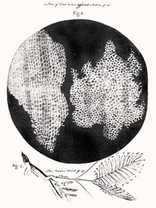
Cells are the fundamental units of life, forming the basis of all living organisms. The development of the microscope in the 1600s enabled scientists to discover cells and single-celled organisms, leading to the formulation of the Cell Theory.
Cell Theory: All living things are composed of one or more cells and their products. All cells arise from pre-existing cells.
Discovery: Robert Hooke named cells after observing cork tissue under a microscope.
Types of Organisms: Organisms can be single-celled (e.g., bacteria, protists) or multicellular (e.g., plants, animals, fungi).
The Microscopic World of Cells
Microscopes have revolutionized biology by allowing the visualization of cells and their structures. There are several types of microscopes, each with unique capabilities.
Light Microscope (LM): Uses visible light to view living or dead specimens up to 2,000x magnification.
Scanning Electron Microscope (SEM): Provides detailed views of cell surfaces; specimens must be killed and coated with metal.
Transmission Electron Microscope (TEM): Reveals internal cell structures at very high magnification; specimens must be killed.
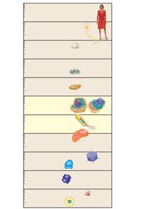
Cell Size and Scale
Cells and their components vary greatly in size, from atoms and small molecules to entire cells visible to the naked eye. Most cells are microscopic, requiring magnification for detailed study.
Types of Cells: Prokaryotic vs. Eukaryotic
Major Domains of Life
All life is classified into three domains: Bacteria, Archaea, and Eukarya. The first two are prokaryotic, while Eukarya includes all eukaryotic organisms (protists, plants, fungi, animals).
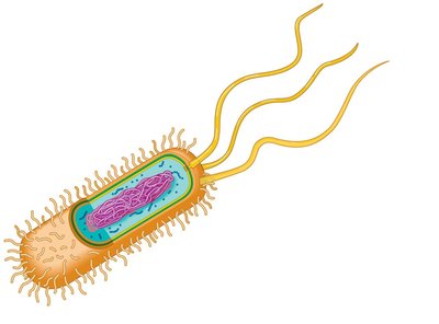
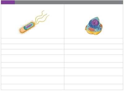
Prokaryotic Cells
Prokaryotic cells are simpler and smaller than eukaryotic cells. They lack membrane-bound organelles and a true nucleus.
Key Features: Plasma membrane, cell wall, capsule, fimbriae, nucleoid (circular DNA), ribosomes, flagella.
Domains: Bacteria and Archaea.
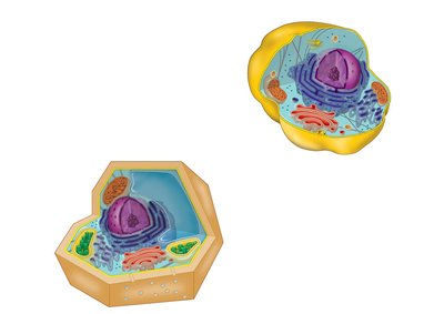
Eukaryotic Cells
Eukaryotic cells are larger and more complex, containing membrane-bound organelles and a nucleus with linear chromosomes.
Key Features: Nucleus, endoplasmic reticulum, Golgi apparatus, mitochondria, cytoskeleton, plasma membrane, and (in plants) chloroplasts, cell wall, central vacuole.
Domains: Eukarya (protists, plants, fungi, animals).
Comparison Table: Prokaryotic vs. Eukaryotic Cells
Prokaryotic Cells | Eukaryotic Cells |
|---|---|
No nucleus (nucleoid region) | True nucleus with nuclear envelope |
No membrane-bound organelles | Membrane-bound organelles present |
Single circular chromosome | Multiple linear chromosomes |
Smaller, simpler | Larger, more complex |
Bacteria, Archaea | Protists, plants, fungi, animals |
Cell Membranes and Structure
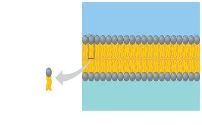
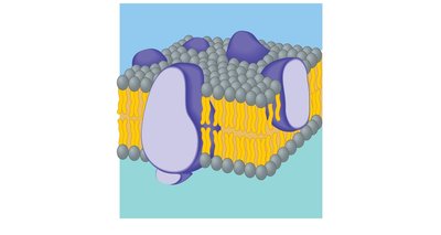
Plasma Membrane Structure
The plasma membrane separates the cell from its environment and regulates the movement of substances. It is composed mainly of a phospholipid bilayer with embedded proteins, forming a fluid mosaic model.
Phospholipids: Amphipathic molecules with hydrophilic heads and hydrophobic tails.
Fluid Mosaic Model: Describes the dynamic arrangement of lipids and proteins.
Cell Walls and Extracellular Structures
Plant Cell Walls and Animal Extracellular Matrix
Plant cells have rigid cell walls made of cellulose, providing protection and structural support. Animal cells lack cell walls but have an extracellular matrix for support and communication.
Cell Junctions: Structures that connect animal cells, allowing coordinated function in tissues.
Genetic Control: The Nucleus and Ribosomes
The Nucleus
The nucleus is the control center of the cell, containing DNA organized into chromosomes. It is surrounded by a double membrane (nuclear envelope) with pores for molecular exchange.
Chromatin: DNA and protein complex forming chromosomes.
Nucleolus: Site of ribosome component synthesis.
Ribosomes
Ribosomes are the sites of protein synthesis. They can be free in the cytoplasm or bound to the endoplasmic reticulum (ER).
Function: Translate messenger RNA (mRNA) into proteins.
Location: Free ribosomes synthesize cytosolic proteins; ER-bound ribosomes synthesize membrane-bound or secreted proteins.
Central Dogma: DNA → RNA → Protein
Genetic information flows from DNA to RNA to protein. DNA is transcribed into mRNA, which is then translated by ribosomes into proteins.
Transcription: DNA → mRNA (in the nucleus)
Translation: mRNA → Protein (in the cytoplasm)
The Endomembrane System
Components and Functions
The endomembrane system includes the nuclear envelope, endoplasmic reticulum (ER), Golgi apparatus, lysosomes, and vacuoles. These organelles work together to manufacture, modify, and transport cellular products.
Endoplasmic Reticulum (ER): Rough ER (with ribosomes) synthesizes proteins; smooth ER synthesizes lipids and detoxifies chemicals.
Golgi Apparatus: Modifies, sorts, and ships proteins and lipids.
Lysosomes: Contain digestive enzymes for recycling macromolecules and organelles.
Vacuoles: Storage and transport; central vacuole in plants maintains turgor pressure.
Energy Conversion: Chloroplasts and Mitochondria
Chloroplasts
Chloroplasts are the sites of photosynthesis in plant and algal cells, converting light energy into chemical energy (sugars).
Structure: Double membrane, thylakoids, stroma.
Function: Photosynthesis:
Mitochondria
Mitochondria are the powerhouses of the cell, converting chemical energy from food into ATP through cellular respiration.
Structure: Double membrane, cristae, matrix.
Function: Cellular respiration:
Endosymbiotic Theory
Chloroplasts and mitochondria contain their own DNA and ribosomes, similar to prokaryotes. This supports the endosymbiotic theory, which proposes that these organelles originated as free-living prokaryotes engulfed by ancestral eukaryotic cells.
The Cytoskeleton and Cell Movement
Cytoskeleton
The cytoskeleton is a network of protein fibers that provides structural support, maintains cell shape, and enables movement.
Microtubules: Hollow tubes for support and movement.
Intermediate Filaments and Microfilaments: Provide additional support and structure.
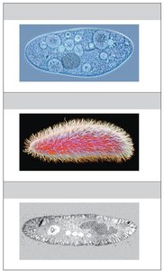
Cilia and Flagella
Cilia and flagella are extensions of the cytoskeleton that aid in cell movement. Flagella are long and few, while cilia are short and numerous.
Flagella: Propel cells with a whip-like motion (e.g., sperm cells).
Cilia: Move in coordinated waves (e.g., lining the respiratory tract).
Evolutionary Connections
Antibiotic Resistance and Natural Selection
Antibiotic resistance in bacteria and resistance to bacterial diseases in humans are examples of evolution by natural selection. Genetic mutations that confer resistance are favored in environments with persistent disease or antibiotic use.
Example: Mutations in Bangladeshi populations confer resistance to cholera.
