 Back
BackA Tour of the Cell: Structure, Function, and Diversity
Study Guide - Smart Notes
 Tailored notes based on your materials, expanded with key definitions, examples, and context.
A Tour of the Cell
Introduction to Cell Biology
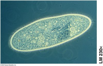
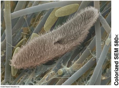
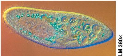
Advancements in microscopy have revolutionized our understanding of cells, the fundamental units of life. Early microscopes were seen as novelties, but pioneers like Robert Hooke and Antonie van Leeuwenhoek used them to discover and name the cell, and to observe diverse cellular structures.
Cell Theory: All living things are composed of cells; all cells arise from pre-existing cells.
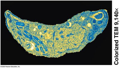
Microscopy: Light microscopes allow observation of living cells, while electron microscopes (SEM and TEM) provide greater magnification and resolution, revealing cellular ultrastructure.
Example: Hooke's observation of cork led to the term "cell"; Leeuwenhoek observed blood, sperm, and pond water organisms.
Microscope Concepts
Magnification: The increase in an object's image size compared to its actual size.
Resolution: The clarity of an image; the ability to distinguish two close objects as separate.
The Cell Surface
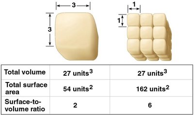
Surface-to-Volume Ratio
The small size of most cells provides a large surface-to-volume ratio, which is critical for efficient exchange of materials with the environment.
Surface Area: Determines the rate of exchange of substances.
Volume: Determines the amount of metabolic activity.
Surface-to-Volume Ratio: Smaller cells have a higher ratio, facilitating efficient transport.
Single Large Cube | 27 Small Cubes | |
|---|---|---|
Total volume | 27 units3 | 27 units3 |
Total surface area | 54 units2 | 162 units2 |
Surface-to-volume ratio | 2 | 6 |
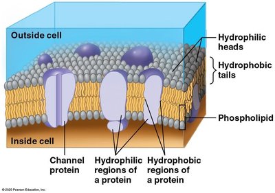
Plasma Membrane Structure
The plasma membrane forms the cell's boundary and regulates the movement of substances. It consists of a phospholipid bilayer with embedded proteins.
Phospholipid Bilayer: Hydrophilic heads face outward; hydrophobic tails face inward.
Channel Proteins: Allow passage of ions and hydrophilic molecules.
Pumps: Use energy to actively transport molecules.
Taxonomic Domains and Cell Types
Classification of Life
Organisms are classified into three domains based on genetic, metabolic, and structural similarities: Bacteria, Archaea, and Eukarya. Eukarya is further divided into four kingdoms: Protista, Plantae, Fungi, and Animalia.
Bacteria: Prokaryotic cells.
Archaea: Prokaryotic cells, biochemically distinct from bacteria.
Eukarya: Eukaryotic cells with membrane-bound organelles.
Prokaryotic vs. Eukaryotic Cells
All cells share basic features: plasma membrane, DNA, ribosomes, and cytosol. Prokaryotic cells (Bacteria and Archaea) are structurally simpler, lacking a membrane-bound nucleus and organelles. Eukaryotic cells (Eukarya) have a nucleus and specialized organelles.
Prokaryotic Cells: Simpler, smaller, no nucleus.
Eukaryotic Cells: Complex, larger, nucleus and organelles.
Prokaryotic Cell Structure
Shapes and Sizes
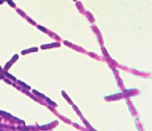
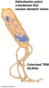
Prokaryotes exhibit three basic shapes: spherical (coccus), rod-shaped (bacillus), and spiral (spirillum or spirochete).
Size Range: 0.2 µm to 750 µm.
Cell Envelope
The cell envelope includes the plasma membrane, cell wall, and glycocalyx. The glycocalyx can be a capsule (regular) or a slime coat (diffuse), providing protection.
Plasma Membrane: Phospholipid bilayer.
Cell Wall: Maintains cell shape.
Glycocalyx: Carbohydrate layer outside the cell wall.

Cytoplasm and Genetic Material
The cytoplasm contains cytosol, nutrients, and cellular machinery. The nucleoid region holds the single circular DNA chromosome, and plasmids are small extrachromosomal DNA rings.
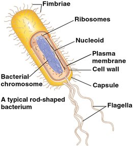
Appendages
Prokaryotic cells may have flagella for motility, fimbriae for adhesion, and conjugation pili for DNA transfer.
Eukaryotic Cell Structure
Compartmentalization
Eukaryotic cells are organized into membrane-enclosed organelles, each performing specific functions. Four basic systems include genetic control, endomembrane system, energy processing, and structural support.
Genetic Control: Nucleus and ribosomes.
Endomembrane System: ER, Golgi apparatus, lysosomes, vacuoles, peroxisomes.
Energy Processing: Mitochondria and chloroplasts.
Structural Support: Cytoskeleton, plasma membrane, cell wall.
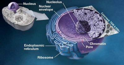
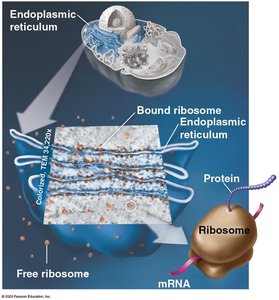
The Nucleus and Ribosomes
The nucleus houses DNA and directs protein synthesis via mRNA. Ribosomes, composed of rRNA and proteins, synthesize proteins according to DNA instructions. Free ribosomes produce cytosolic proteins; bound ribosomes (on rough ER) produce proteins for membranes or export.
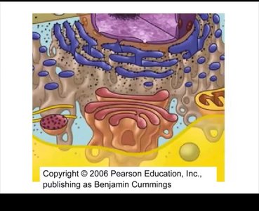
The Endomembrane System
The endomembrane system includes the nuclear envelope, ER, Golgi apparatus, vesicles, lysosomes, and vacuoles. It is involved in synthesis, distribution, storage, and export of cellular products.
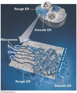
Endoplasmic Reticulum (ER)
The ER is a network of tubes and sacs continuous with the nuclear envelope. The smooth ER synthesizes lipids and processes toxins; the rough ER produces membranes and secretory proteins.
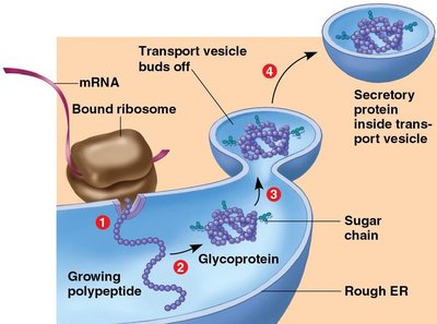
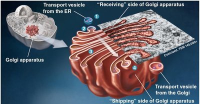
Golgi Apparatus
The Golgi apparatus modifies, sorts, and ships cell products received from the ER. It consists of stacks of membranous sacs.
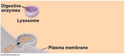
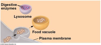
Lysosomes
Lysosomes are membrane-bound vesicles containing digestive enzymes. They break down biomolecules and recycle cellular components.
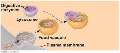
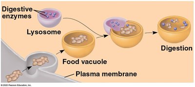
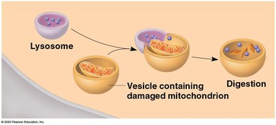
Lysosome Formation
Peroxisomes
Peroxisomes are membrane-bound vesicles with enzymes that catalyze reactions producing hydrogen peroxide. They are active in lipid metabolism and detoxification.
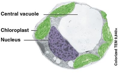
Vacuoles
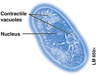
Vacuoles are large vesicles with diverse functions. Plant cells have a central vacuole for storage and growth; some protists have contractile vacuoles for osmoregulation.
Review of the Endomembrane System
Transport vesicles connect the endomembrane system, carrying proteins and lipids from the ER to the Golgi, where they are modified and sorted, then sent to their destination or exported.
Energy-Converting Organelles
Mitochondria
Mitochondria are found in almost all eukaryotes. They are endosymbionts with their own genome and ribosomes, surrounded by a double membrane. The inner membrane is highly folded, increasing surface area for cellular respiration and ATP production.
Chloroplasts
Chloroplasts are found in plants and algae. They have their own genome and multiple membranes, including thylakoid disks containing chlorophyll for photosynthesis.
Endosymbiotic Theory
The endosymbiotic theory explains the origin of mitochondria and chloroplasts as formerly independent prokaryotes engulfed by ancestral eukaryotic cells. Evidence includes their bacterial-like DNA, ribosomes, double membranes, and independent reproduction.
The Cytoskeleton and Cell Surfaces
Cytoskeleton
The cytoskeleton is an internal network of protein filaments (microfilaments, intermediate filaments, microtubules) that maintains cell shape, enables movement, and anchors organelles.
Cilia and Flagella
Eukaryotic cilia and flagella are locomotor appendages made of microtubules in a "9 + 2" arrangement. Flagella are longer and move with a whiplike motion; cilia are shorter and move in coordinated waves.
Extracellular Matrix (ECM)
Animal cells secrete an ECM that binds cells, supports the plasma membrane, and communicates with the cytoskeleton. ECM attaches to cells via glycoproteins and integrins.
Cell Junctions
Animal tissues have specialized junctions: tight junctions (seal cells), anchoring junctions (strengthen tissues), and gap junctions (allow communication).
Cell Walls and Plasmodesmata
Plant cells have rigid cell walls composed of cellulose, providing support and protection. Plasmodesmata are channels between cells for sharing water, nutrients, and signals.
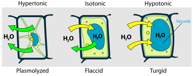
Turgor Pressure in Plant Cells
Turgor pressure is the force exerted by water inside the central vacuole against the cell wall. It is essential for maintaining plant rigidity and growth. Water movement depends on the tonicity of the environment.
Hypertonic: Water leaves the cell, causing plasmolysis.
Isotonic: No net water movement; cell is flaccid.
Hypotonic: Water enters the cell, making it turgid.
Additional info: Academic context was added to clarify the functions and relationships of cell structures, the endomembrane system, and the significance of surface-to-volume ratio.
