 Back
BackA Tour of the Cell: Structure, Function, and Evolution
Study Guide - Smart Notes
 Tailored notes based on your materials, expanded with key definitions, examples, and context.
A Tour of the Cell
Cell Theory and the Fundamental Unit of Life
The cell is the simplest collection of matter that can be alive, forming the foundation of all living organisms. Cell theory states that all organisms are made of cells and all cells arise from pre-existing cells, establishing a universal lineage among cells.
Cell Theory: All living things are composed of cells; cells are the basic unit of structure and function; new cells arise from existing cells.
Common Features: Despite diversity, all cells share certain structural features.
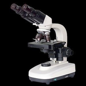
Microscopy: Observing Cells
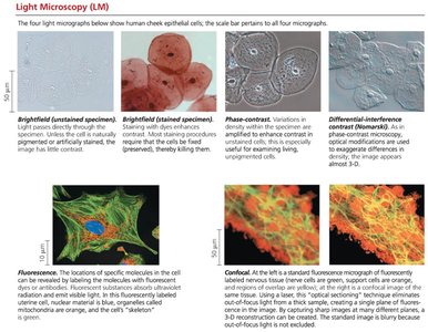
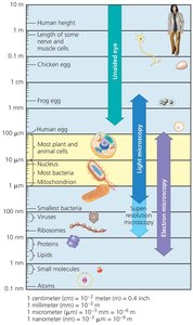
Microscopes are essential tools for studying cells, which are too small to be seen with the naked eye. Light microscopes (LM) use visible light and glass lenses to magnify specimens, while electron microscopes (EM) use electron beams for higher resolution.
Magnification: Ratio of image size to actual size.
Resolution: Minimum distance between two distinguishable points.
Contrast: Difference in brightness between areas of the image.
Light Microscopes: Can magnify up to 1,000x; advanced techniques include fluorescence and confocal microscopy.
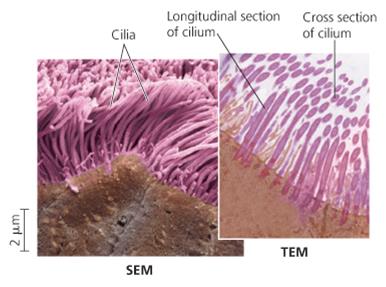
Electron Microscopes: SEMs provide 3D surface images; TEMs reveal internal structures.
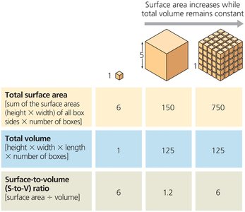
Cell Size and Surface Area-to-Volume Ratio
Cell size is constrained by metabolic requirements. The surface area-to-volume ratio is critical for efficient exchange of materials.
Surface Area: Increases with the square of the cell's linear dimension.
Volume: Increases with the cube of the cell's linear dimension.
Small Cells: Have a higher surface area-to-volume ratio, supporting efficient metabolism.
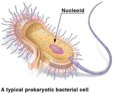
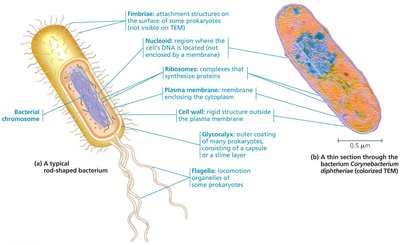
Prokaryotic and Eukaryotic Cells
Prokaryotic Cells
Prokaryotic cells are small, simple, and lack membrane-bound organelles. They include the domains Bacteria and Archaea.
Characteristics: No nucleus; DNA in nucleoid; no organelles; unicellular.
First Appeared: 3.5 billion years ago.
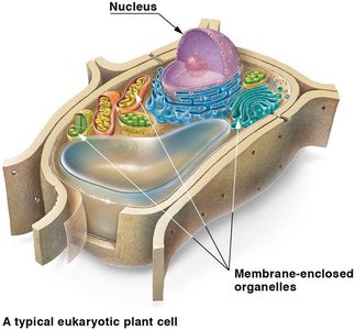
Eukaryotic Cells
Eukaryotic cells are larger and more complex, with membrane-enclosed organelles and a nucleus. They include protists, fungi, animals, and plants.
Characteristics: DNA in nucleus; organelles; unicellular or multicellular.
First Appeared: 2.1 billion years ago.
Basic Features of All Cells
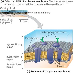
Plasma Membrane: Selective barrier for exchange of materials.
Cytosol: Semifluid substance inside the cell.
Chromosomes: Carry genetic information.
Ribosomes: Synthesize proteins.
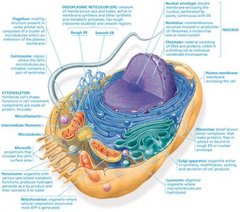
Internal Organization of Eukaryotic Cells
Nucleus: Information Central
The nucleus houses most of the cell's DNA and is surrounded by a double membrane called the nuclear envelope. The nucleolus within the nucleus is the site of ribosomal RNA synthesis.
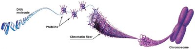
Chromosomes: DNA molecules associated with proteins.
Chromatin: Complex of DNA and proteins; condenses during cell division.
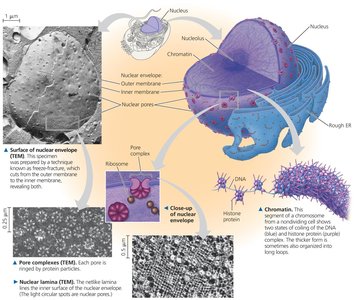
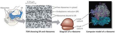
Ribosomes: Protein Factories
Ribosomes are complexes of ribosomal RNA and protein, responsible for protein synthesis. They can be free in the cytosol or bound to the endoplasmic reticulum or nuclear envelope.
Free Ribosomes: Synthesize proteins for use within the cell.
Bound Ribosomes: Synthesize proteins for secretion or membrane insertion.
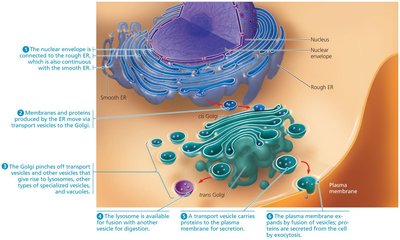
The Endomembrane System
The endomembrane system regulates protein traffic and performs metabolic functions. It includes the nuclear envelope, endoplasmic reticulum, Golgi apparatus, lysosomes, vacuoles, and plasma membrane.
Components: Connected directly or via vesicles.
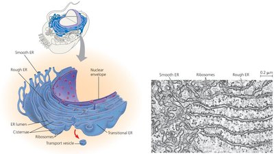
Endoplasmic Reticulum (ER)
The ER is a biosynthetic factory, continuous with the nuclear envelope, and consists of two regions: smooth ER (lacks ribosomes) and rough ER (studded with ribosomes).
Smooth ER: Synthesizes lipids, metabolizes carbohydrates, detoxifies drugs, stores calcium ions.
Rough ER: Produces glycoproteins, distributes transport vesicles, manufactures membranes.
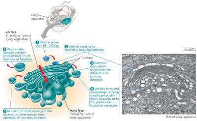
Golgi Apparatus
The Golgi apparatus consists of flattened sacs called cisternae and functions as the cell's shipping and receiving center.
Functions: Modifies ER products, manufactures macromolecules, sorts and packages materials into vesicles.
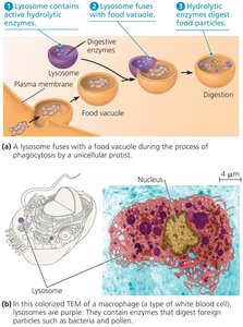
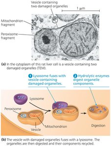
Lysosomes
Lysosomes are membranous sacs of hydrolytic enzymes that digest macromolecules. They are involved in phagocytosis and autophagy.
Phagocytosis: Engulfing and digesting other cells or particles.
Autophagy: Recycling the cell's own organelles and macromolecules.
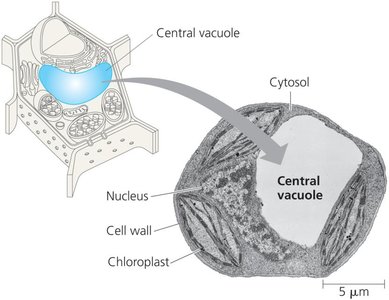
Vacuoles
Vacuoles are large vesicles derived from the ER and Golgi apparatus, with diverse functions in different cell types.
Food Vacuoles: Formed by phagocytosis.
Contractile Vacuoles: Pump excess water out of cells.
Central Vacuole: In plant cells, stores inorganic ions and maintains cell structure.
Relationships Among Organelles
Organelles of the endomembrane system interact through vesicle transport, coordinating cellular functions.
Energy Conversion: Mitochondria and Chloroplasts
Mitochondria
Mitochondria are the sites of cellular respiration, converting oxygen and nutrients into ATP. They have a double membrane, with the inner membrane folded into cristae.
Compartments: Intermembrane space and mitochondrial matrix.
Cristae: Increase surface area for ATP synthesis.
Chloroplasts
Chloroplasts are found in plants and algae, and are the sites of photosynthesis. They contain chlorophyll, thylakoids (stacked as grana), and stroma (internal fluid).
Thylakoids: Membranous sacs for light reactions.
Stroma: Site of the Calvin cycle.
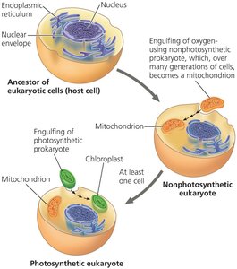
Endosymbiont Theory
The endosymbiont theory proposes that mitochondria and chloroplasts originated from prokaryotic cells engulfed by ancestral eukaryotes. These organelles share similarities with bacteria, such as double membranes, circular DNA, and ribosomes.
Other Organelles
Peroxisomes
Peroxisomes are specialized metabolic compartments that produce hydrogen peroxide and convert it to water. They detoxify harmful substances and perform various metabolic reactions.
Cytoskeleton: Structure and Motility
Cytoskeleton Overview
The cytoskeleton is a network of fibers that organizes cell structures and activities, providing support, shape, and motility.
Microtubules: Thickest fibers; shape, support, organelle movement, chromosome separation.
Microfilaments (Actin Filaments): Thinnest; bear tension, support, motility (muscle contraction, cytoplasmic streaming).
Intermediate Filaments: Middle diameter; reinforce shape, anchor organelles, more permanent.
Microtubules: Centrosomes, Cilia, and Flagella
Microtubules grow from centrosomes and are involved in the movement of cilia and flagella. Cilia move with a back-and-forth motion, while flagella move in a whip-like manner.
Centrosome: Microtubule-organizing center with centrioles.
Cilia and Flagella: Extensions for cell movement; powered by dynein motor proteins.
Microfilaments and Intermediate Filaments
Microfilaments are built from actin and interact with myosin for cellular movement. Intermediate filaments provide structural stability and are more permanent than other cytoskeletal elements.
Extracellular Components and Cell Junctions
Cell Walls of Plants
Plant cells have cell walls made of cellulose, providing protection, shape, and preventing excessive water uptake. Cell walls may have multiple layers: primary wall, middle lamella, and secondary wall.
Extracellular Matrix (ECM) of Animal Cells
Animal cells lack cell walls but have an extracellular matrix composed of glycoproteins like collagen, proteoglycans, and fibronectin. ECM proteins bind to integrins in the plasma membrane, facilitating cell communication and structural support.
Cell Junctions
Cells adhere, interact, and communicate through specialized junctions:
Plasmodesmata: Channels between plant cells for transport of water, solutes, proteins, and RNA.
Tight Junctions: Seal animal cells together, preventing leakage.
Desmosomes: Anchor cells together like rivets.
Gap Junctions: Channels for molecular signals and nutrients between animal cells.
