 Back
BackA Tour of the Cell: Structure and Function in Eukaryotic and Prokaryotic Cells
Study Guide - Smart Notes
 Tailored notes based on your materials, expanded with key definitions, examples, and context.
Introduction to the Cell
Microscopes Reveal the World of the Cell
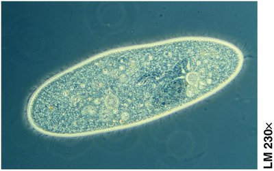
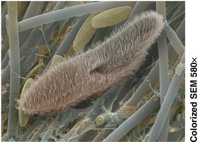
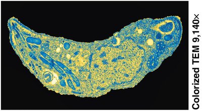
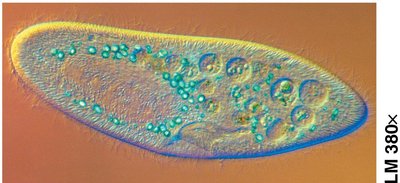
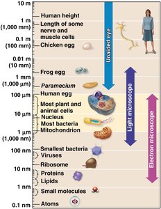
Microscopes are essential tools for studying cell structure and function. The light microscope allows observation of living cells, while electron microscopes (scanning and transmission) provide greater magnification and resolution, revealing cellular ultrastructure. Magnification is the increase in an object's image size compared to its actual size, and resolution is the clarity of an image, or the ability to distinguish two nearby objects as separate.
Cell theory: All living things are composed of cells, and all cells come from other cells.
Microscopy has enabled the discovery and understanding of cell structure and function.
Cell Size and the Plasma Membrane
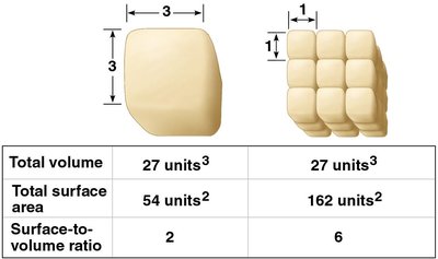
The Small Size of Cells Relates to the Need to Exchange Materials
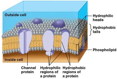
Most cells are microscopic, which provides a large surface-to-volume ratio. This ratio is crucial for efficient exchange of materials across the plasma membrane. The plasma membrane is a phospholipid bilayer with embedded proteins, some forming channels for ions and hydrophilic molecules, others acting as pumps for active transport.
Surface-to-volume ratio limits cell size; smaller cells have more surface area relative to their volume.
Membrane proteins facilitate selective transport and communication.
Prokaryotic and Eukaryotic Cells
Prokaryotic Cells Are Structurally Simpler
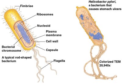
All cells share basic features: plasma membrane, DNA, ribosomes, and cytosol. Prokaryotic cells (Bacteria and Archaea) are smaller and simpler, lacking a membrane-bound nucleus and organelles. Their DNA is located in the nucleoid region, and they may have flagella for movement.
Prokaryotic cells: No nucleus, no membrane-bound organelles.
Eukaryotic cells: Membrane-enclosed nucleus and organelles.
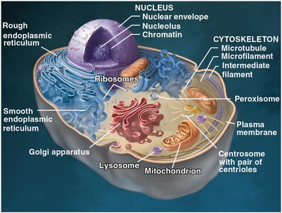
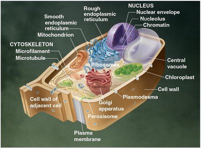
Eukaryotic Cells Are Partitioned into Functional Compartments
Eukaryotic cells (domain Eukarya) are characterized by compartmentalization via membrane-enclosed organelles. These organelles perform specialized functions and are organized into four functional groups:
Genetic control: Nucleus and ribosomes
Manufacture, distribution, breakdown: Endoplasmic reticulum, Golgi apparatus, lysosomes, vacuoles, peroxisomes
Energy processing: Mitochondria (all cells), chloroplasts (plant cells)
Structural support, movement, communication: Cytoskeleton, plasma membrane, cell wall (plants)
The Nucleus and Ribosomes
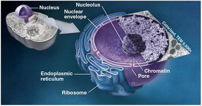
The Nucleus Contains the Cell’s Genetic Instructions
The nucleus houses DNA, which directs protein synthesis via messenger RNA (mRNA). The nucleolus is the site of ribosome subunit assembly.
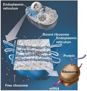
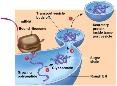
Ribosomes Make Proteins for Use in the Cell and Export
Ribosomes are composed of ribosomal RNA (rRNA) and proteins. They synthesize proteins according to instructions from DNA. Cells with high protein production have many ribosomes.
The Endomembrane System
Many Organelles Are Connected in the Endomembrane System
The endomembrane system includes organelles that interact in the synthesis, distribution, storage, and export of molecules. These organelles are structurally and functionally interconnected.
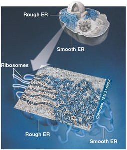
The Endoplasmic Reticulum (ER) is a Biosynthetic Workshop
The ER is a network of tubes and sacs. Smooth ER synthesizes lipids and processes toxins. Rough ER produces membranes, and its ribosomes make membrane and secretory proteins.
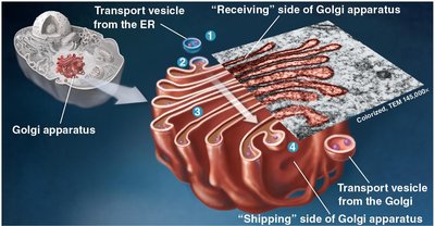
The Golgi Apparatus Modifies, Sorts, and Ships Cell Products
The Golgi apparatus consists of stacks of sacs where products from the ER are processed and sent to other organelles or the cell surface.
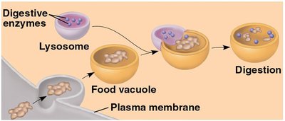
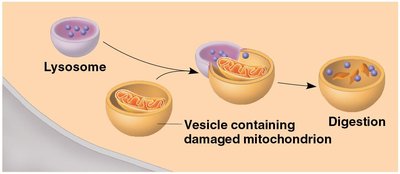
Lysosomes Are Digestive Compartments Within a Cell
Lysosomes contain enzymes that break down ingested substances and damaged organelles, facilitating recycling within the cell.
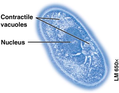
Vacuoles Function in the General Maintenance of the Cell
Vacuoles are large vesicles with diverse functions. Protists have contractile vacuoles for expelling excess water, while plant cells have a central vacuole for storage and growth.
Energy-Converting Organelles
Mitochondria Harvest Chemical Energy from Food
Mitochondria are organelles that carry out cellular respiration in eukaryotic cells. They have two internal compartments: the intermembrane space and the mitochondrial matrix, which contains DNA, ribosomes, and enzymes for respiration.
Chloroplasts Convert Solar Energy to Chemical Energy
Chloroplasts are the photosynthesizing organelles of plants and algae, converting light energy into the chemical energy of sugars.
Evolution Connection: Mitochondria and Chloroplasts Evolved by Endosymbiosis
The endosymbiont theory proposes that mitochondria and chloroplasts were once small prokaryotes that began living within larger cells, explaining their similarities to prokaryotes.
The Cytoskeleton and Cell Surfaces
The Cell’s Internal Skeleton Helps Organize Its Structure and Activities
The cytoskeleton is composed of microfilaments, intermediate filaments, and microtubules. These fibers maintain cell shape, anchor and move organelles, enable amoeboid movement, and facilitate muscle contraction.
Cilia and Flagella Move When Microtubules Bend
Eukaryotic cilia and flagella are locomotor appendages made of microtubules in a "9 + 2" arrangement. Flagella propel cells with a whiplike motion, while cilia move in a coordinated, oar-like fashion.
The Extracellular Matrix of Animal Cells Functions in Support and Regulation
Animal cells synthesize and secrete an extracellular matrix (ECM) that binds tissue cells, supports the plasma membrane, and communicates with the cytoskeleton. The ECM attaches to cells via glycoproteins and integrins.
Three Types of Cell Junctions Are Found in Animal Tissues
Specialized junctions allow cells to adhere, interact, and communicate:
Tight junctions: Bind cells to form leakproof sheets.
Anchoring junctions: Rivet cells into strong tissues.
Gap junctions: Allow ions and small molecules to flow between cells.
Cell Walls Enclose and Support Plant Cells
Plant cells have a rigid cell wall composed primarily of cellulose, providing protection and structural support. Plasmodesmata are cell junctions that allow plant tissues to share water, nutrients, and chemical messages.
Review: Eukaryotic Cell Structures and Their Functions
Functional Categories of Organelles
Eukaryotic cell structures can be grouped by function:
Genetic control
Manufacturing, distribution, breakdown
Energy processing
Structural support, movement, communication
Table: Eukaryotic Cell Structures and Their Functions
Organelle | Main Function |
|---|---|
Nucleus | Genetic control |
Ribosomes | Protein synthesis |
Endoplasmic Reticulum | Synthesis and processing of proteins/lipids |
Golgi Apparatus | Modification, sorting, shipping of cell products |
Lysosomes | Digestion and recycling |
Vacuoles | Storage and maintenance |
Mitochondria | Energy conversion (cellular respiration) |
Chloroplasts | Energy conversion (photosynthesis) |
Cytoskeleton | Structural support, movement |
Plasma Membrane | Selective transport, communication |
Cell Wall (plants) | Protection, support |
Metric Measurement Equivalents
Understanding cell size requires knowledge of metric units:
1 meter (m) = 100 centimeters (cm) = 1,000 millimeters (mm)
1 centimeter (cm) = 0.01 m
1 millimeter (mm) = 0.001 m
1 micrometer (μm) = 0.000001 m
1 nanometer (nm) = 0.000000001 m
Additional info: The notes above expand on brief points with academic context, definitions, and examples to ensure completeness and clarity for exam preparation.
