 Back
BackA Tour of the Cell: Structure, Function, and Organization
Study Guide - Smart Notes
 Tailored notes based on your materials, expanded with key definitions, examples, and context.
Microscopy and Cell Visualization
Microscopy Parameters and Types
Microscopy is essential for studying cell structure and function. Three key parameters define microscopy:
Magnification: The ratio of an object's image size to its real size.
Resolution: The clarity of the image, or the minimum distance between two distinguishable points.
Contrast: Visible differences in brightness between parts of the sample.
Light microscopes (LM) use visible light and glass lenses to magnify specimens, but their resolution is limited for studying organelles. Electron microscopes (EM) provide higher resolution:
Scanning Electron Microscope (SEM): Produces 3-D images of specimen surfaces.
Transmission Electron Microscope (TEM): Reveals internal cell structures.
Recent advances include fluorescent labeling, confocal microscopy, and cryo-electron microscopy, which allow detailed visualization of cellular structures.
Cell Fractionation
Cell fractionation separates organelles using centrifugation, enabling scientists to study their functions. This technique correlates cell structure with biochemical activity.
Cell Types and Comparative Structure
Prokaryotic vs. Eukaryotic Cells
All living organisms are composed of cells, which are classified as prokaryotic or eukaryotic:
Prokaryotic Cells: Found in Bacteria and Archaea. Characterized by the absence of a nucleus and membrane-bound organelles. DNA is located in the nucleoid region.
Eukaryotic Cells: Found in protists, fungi, animals, and plants. Characterized by a nucleus enclosed by a double membrane and numerous membrane-bound organelles. Generally larger than prokaryotic cells.
All cells share basic features: plasma membrane, cytosol, chromosomes, and ribosomes.
Surface Area to Volume Ratio
The plasma membrane regulates the passage of substances. As cells grow, their volume increases faster than surface area, limiting cell size. Cells adapt by dividing, changing shape, or developing extensions to increase surface area.
Cell Diversity and Specialization
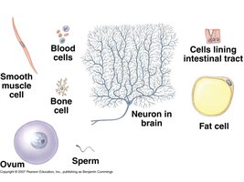
Eukaryotic cells are specialized for different functions, reflected in their unique structures.
Examples: Neurons, blood cells, smooth muscle cells, bone cells, fat cells, ovum, sperm, and cells lining the intestinal tract.
Internal Membranes and Organelles
The Nucleus
The nucleus is the cell's information center, containing most genetic material. It is surrounded by a double-membrane nuclear envelope with nuclear pores for molecular transport. The nuclear lamina and matrix provide structural support. DNA is organized as chromatin, which condenses into chromosomes during cell division. The nucleolus synthesizes ribosomal RNA (rRNA).
Ribosomes
Ribosomes are complexes of rRNA and protein, responsible for protein synthesis. They are found free in the cytosol or bound to the endoplasmic reticulum (ER) or nuclear envelope.
The Endomembrane System
The endomembrane system includes the nuclear envelope, ER, Golgi apparatus, lysosomes, peroxisomes, vacuoles, and plasma membrane. These components are interconnected and regulate protein traffic and metabolic functions.
Endoplasmic Reticulum (ER)
Smooth ER: Synthesizes lipids, detoxifies drugs, stores calcium ions, and metabolizes carbohydrates.
Rough ER: Studded with ribosomes; synthesizes glycoproteins, distributes transport vesicles, and produces membranes.
Golgi Apparatus
Consists of cisternae; modifies ER products, manufactures macromolecules, and sorts/packages materials into vesicles.
Lysosomes
Membranous sacs containing hydrolytic enzymes for digesting macromolecules. They participate in phagocytosis and autophagy.
Peroxisomes
Oxidative organelles that break down fatty acids and detoxify harmful substances. Contain catalase to decompose hydrogen peroxide.
Vacuoles
Food vacuoles: Formed by phagocytosis.
Contractile vacuoles: Pump excess water out of cells.
Central vacuole: Stores ions and supports plant cell growth.
Energy Conversion Organelles
Mitochondria and Chloroplasts
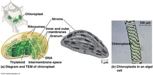
Mitochondria are the site of cellular respiration, generating ATP. Chloroplasts, found in plants and algae, conduct photosynthesis. Both organelles are semiautonomous and share similarities with bacteria, supporting the endosymbiont theory.
Mitochondria: Double membrane, cristae, matrix, own DNA and ribosomes.
Chloroplasts: Double membrane, thylakoids (stacked as grana), stroma, own DNA and ribosomes.
The Cytoskeleton
Components and Functions
The cytoskeleton provides structural support, maintains cell shape, and enables motility. It consists of:
Microtubules: Hollow rods made of tubulin; shape the cell, guide organelle movement, and separate chromosomes.
Microfilaments (Actin Filaments): Twisted chains of actin; support cell shape, form microvilli, and enable motility.
Intermediate Filaments: More permanent structures; support cell shape and anchor organelles.
Specialized Structures
Centrosomes and Centrioles: Organize microtubules in animal cells.
Cilia and Flagella: Microtubule-based extensions for movement; powered by dynein motor proteins.
Cytoplasmic Streaming: Actin-driven flow of cytoplasm in plant cells.
Extracellular Components and Cell Connections
Cell Walls
Plant cells have cell walls made of cellulose, providing protection, shape, and preventing excessive water uptake. Cell walls may have primary, middle lamella, and secondary layers.
Extracellular Matrix (ECM) in Animal Cells
Animal cells are surrounded by ECM composed of glycoproteins (collagen, proteoglycans, fibronectin). ECM interacts with integrins in the plasma membrane, influencing cell behavior and gene activity.
Cell Junctions
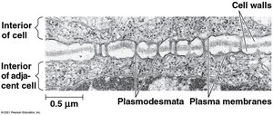
Plasmodesmata: Channels connecting plant cells, allowing passage of water, solutes, proteins, and RNA.
Tight Junctions: Seal neighboring animal cells to prevent leakage.
Desmosomes: Anchor cells together into strong sheets.
Gap Junctions: Provide cytoplasmic channels for communication between animal cells.
Integration of Cellular Components
Coordination of Cell Activities
Cellular functions are coordinated by the interaction of various components. For example, macrophages use the cytoskeleton, lysosomes, and plasma membrane to destroy bacteria, demonstrating the integrated nature of cell structure and function.
Summary Table: Cytoskeletal Elements
The following table summarizes the main cytoskeletal elements and their properties:
Element | Diameter | Composition | Main Functions |
|---|---|---|---|
Microtubules | ~25 nm | Tubulin dimers | Cell shape, organelle movement, chromosome separation |
Microfilaments | ~7 nm | Actin | Cell shape, muscle contraction, cell motility |
Intermediate Filaments | 8–12 nm | Various proteins (e.g., keratin) | Cell shape, organelle anchoring |
Key Equations
Surface area to volume ratio for a spherical cell:
This ratio decreases as cell size increases, affecting transport efficiency.
