 Back
A Tour of the Cell: Structure, Function, and Diversity
Study Guide - Smart Notes
 Tailored notes based on your materials, expanded with key definitions, examples, and context.
Ch. 6 - A Tour of the Cell
Introduction to Cell Biology
Cells are the fundamental units of life, and their study is central to understanding biology. This chapter explores the structure, diversity, and function of cells, focusing on the differences between prokaryotic and eukaryotic cells, the use of microscopes, and the organization of cellular components.
Microscopes and Cell Visualization
Types of Microscopes
Microscopes are essential tools for visualizing cells and their structures, which are often too small to be seen with the naked eye.
Light Microscopes: Use visible light to magnify objects up to about 1000x, allowing observation of living cells and some organelles.
Electron Microscopes: Use beams of electrons for much higher magnification and resolution, revealing detailed cell structures.
Types of Electron Microscopes:
Scanning Electron Microscope (SEM): Visualizes external cell surfaces in high detail.
Transmission Electron Microscope (TEM): Visualizes internal cell structures by passing electrons through thin sections of specimens.
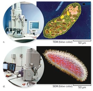
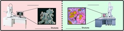
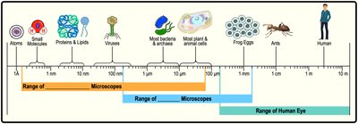
Example: To measure the size of a ribosome inside a eukaryotic cell, a transmission electron microscope (TEM) would be used.
Prokaryotic and Eukaryotic Cells
Major Cell Types
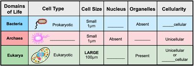
All life is classified into two broad cell types: prokaryotic and eukaryotic cells.
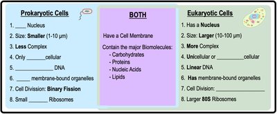
Prokaryotic Cells: Lack a nucleus and membrane-bound organelles. Includes domains Bacteria and Archaea.
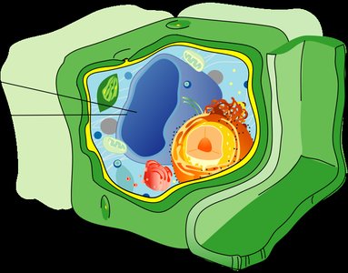
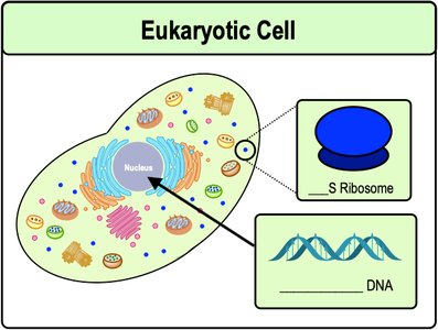
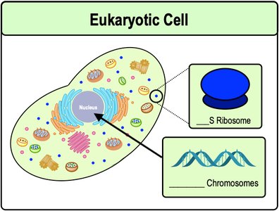
Eukaryotic Cells: Possess a nucleus and various membrane-bound organelles. Includes domain Eukarya (plants, animals, fungi, protists).
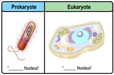
Features of Bacterial (Prokaryotic) Cells
Bacteria: The most abundant and diverse organisms on Earth.
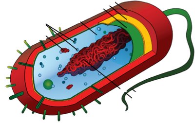
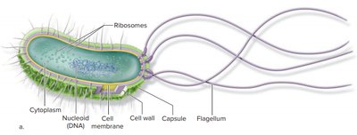
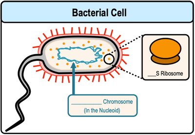
DNA: Circular in shape, located in a region called the nucleoid (not membrane-bound).
Ribosomes: Small (70S), responsible for protein synthesis.
Cell Division: Occurs by binary fission.
Features of Eukaryotic Cells
Nucleus: Contains linear DNA and is surrounded by a nuclear envelope.
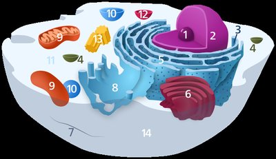
Organelles: Includes mitochondria, endoplasmic reticulum, Golgi apparatus, lysosomes, etc.
Ribosomes: Larger (80S) than those in prokaryotes.
Cell Division: Occurs by mitosis and cytokinesis.
Comparison of Prokaryotic and Eukaryotic Cells
Key differences include cell size, presence of a nucleus, organelles, ribosome size, and modes of motility.
Feature | Prokaryotic Cells | Eukaryotic Cells |
|---|---|---|
Nucleus | Absent | Present |
Organelles | Absent | Present |
DNA | Circular | Linear |
Ribosomes | 70S | 80S |
Cell Size | 1-10 μm | 10-100 μm |
Cell Division | Binary Fission | Mitosis/Cytokinesis |
Introduction to Eukaryotic Organelles
Overview of Organelles
Eukaryotic cells contain several membrane-bound organelles, each with specialized functions. Some organelles are unique to plant or animal cells.
Ribosomes
Ribosomes: Non-membranous molecular machines that synthesize proteins in all living cells.
Protein Synthesis (Translation): The process by which ribosomes build proteins from mRNA templates.
Ribosomes can be free in the cytoplasm or bound to the rough endoplasmic reticulum (ER).
The Endomembrane System
Components and Functions
The endomembrane system is a group of membrane-bound organelles within eukaryotic cells that work together in the synthesis, modification, and transport of proteins and lipids.
Organelles are interconnected by vesicles (small membrane bubbles).
Functions include protein secretion, cellular digestion, and recycling.
Protein Secretion Pathway
Nucleus: Stores DNA, which is transcribed into RNA. The nucleolus within the nucleus assembles ribosomes.
Endoplasmic Reticulum (ER):
Rough ER (rER): Studded with ribosomes; site of protein folding and modification.
Smooth ER (sER): Lacks ribosomes; synthesizes lipids and detoxifies drugs/poisons.
Golgi Apparatus: Receives proteins and lipids from the ER, modifies, sorts, and packages them into vesicles for export.
Example Pathway: Rough ER → Golgi Apparatus → Transport Vesicle → Cell Membrane (for secretion)
Digestive Organelles
Lysosomes: Acidic vesicles containing digestive enzymes; break down food, debris, and bacteria. Primarily in animal cells.
Peroxisomes: Vesicles with enzymes that break down hydrogen peroxide and fatty acids; found in all eukaryotic cells.
Central Vacuole: Large vesicle in plant cells for storage, degradation, and maintaining turgor pressure.
Mitochondria and Chloroplasts
Mitochondria
Function: Synthesize ATP (adenosine triphosphate) via cellular respiration, providing energy for the cell.
Structure: Double membrane (outer and highly folded inner membrane called cristae), intermembrane space, and matrix (contains mitochondrial DNA and ribosomes).
Chloroplasts
Function: Site of photosynthesis in plant cells, converting solar energy into chemical energy (glucose).
Structure: Double membrane, thylakoids (disk-shaped sacs), grana (stacks of thylakoids), and stroma (fluid containing DNA and ribosomes).
Endosymbiotic Theory
The endosymbiotic theory proposes that mitochondria and chloroplasts originated as free-living prokaryotes engulfed by ancestral eukaryotic cells, forming a symbiotic relationship.
Evidence: Both organelles have circular DNA, 70S ribosomes, replicate by binary fission, and possess double membranes.
The Cytoskeleton
Components and Functions
The cytoskeleton is a network of protein filaments that provides cell shape, structure, movement, and intracellular transport.
Microfilaments: Thinnest, made of actin; involved in cell movement and shape.
Intermediate Filaments: Medium thickness; provide mechanical support.
Microtubules: Thickest; made of tubulin, form tracks for vesicle transport and are structural components of cilia and flagella.
Cilia and Flagella: Microtubule-based structures for cell movement. Cilia are short and numerous; flagella are long and few.
Cell Junctions
Cell junctions are specialized structures that connect adjacent cells, allowing communication and maintaining tissue integrity.
Tight Junctions: Create impermeable barriers between cells.
Anchoring Junctions (Desmosomes): Provide strong adhesion between cells.
Gap Junctions: Protein channels that allow exchange of small molecules and ions between animal cells.
Plasmodesmata: Channels connecting the cytoplasm of adjacent plant cells.
