 Back
A Tour of the Cell: Structure, Function, and Organization
Study Guide - Smart Notes
 Tailored notes based on your materials, expanded with key definitions, examples, and context.
The Fundamental Units of Life
Introduction to Cells
All living organisms are composed of cells, which are the simplest units of life. Cells share common features but can differ greatly in structure and function.
Cell Theory: All organisms are made of cells; all cells arise from pre-existing cells.
Cell Diversity: Cells vary in size, shape, and function, but all have basic structural components.
Microscopy
Types and Parameters of Microscopy
Microscopes are essential tools for visualizing cells, which are typically too small to be seen with the naked eye.
Light Microscopes (LM): Use visible light and glass lenses to magnify specimens.
Magnification: Ratio of image size to actual size.
Resolution: Clarity of the image; minimum distance between two distinguishable points.
Contrast: Differences in brightness between parts of the sample.
Microscopy Techniques
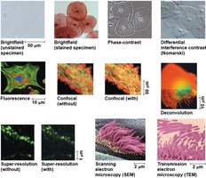
Various microscopy techniques enhance contrast and resolution, allowing detailed study of cell structures.
Electron Microscopes (EM): Use electron beams for higher resolution.
Scanning Electron Microscope (SEM): Provides 3D images of cell surfaces.
Transmission Electron Microscope (TEM): Reveals internal cell structures.
Fluorescence, Confocal, and Super-resolution Microscopy: Allow visualization of specific cell components and finer details.
Cell Size and Scale
Size Range of Cells
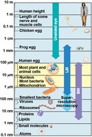
Cells and their components span a wide range of sizes, from atoms to entire organisms.
Most plant and animal cells: 10–100 μm.
Bacteria: 1–10 μm.
Organelles: Smaller than cells, visible only with advanced microscopy.
Cell Fractionation
Separation of Cell Components
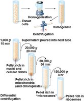
Cell fractionation is a technique used to isolate and study individual organelles.
Centrifugation: Separates cell components based on size and density.
Biochemistry and Cytology: Correlate cell structure with function.
Prokaryotic vs. Eukaryotic Cells
Basic Features of All Cells
All cells share certain features:
Plasma membrane: Selective barrier for transport.
Cytosol: Semifluid substance inside the cell.
Chromosomes: Carry genetic information.
Ribosomes: Synthesize proteins.
Prokaryotic Cells
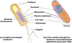
Prokaryotes (Bacteria and Archaea) lack a nucleus and membrane-bound organelles.
Nucleoid: Region where DNA is located.
Cytoplasm: Enclosed by plasma membrane.
Eukaryotic Cells
Eukaryotes (Protists, Fungi, Animals, Plants) have a nucleus and membrane-bound organelles.
Nucleus: Enclosed by nuclear envelope.
Organelles: Specialized compartments for cellular functions.
Cytoplasm: Region between plasma membrane and nucleus.
Plasma Membrane Structure
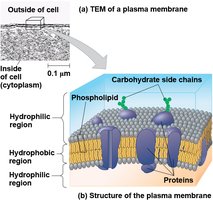
The plasma membrane is a phospholipid bilayer with embedded proteins, regulating transport.
Hydrophilic and Hydrophobic Regions: Allow selective permeability.
Cell Size and Surface Area
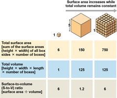
Cell size is limited by the surface area-to-volume ratio, which affects metabolic efficiency.
As cell size increases: Volume grows faster than surface area.
Surface-to-volume ratio: Critical for nutrient and waste exchange.
Organization of Eukaryotic Cells
Animal and Plant Cell Structure
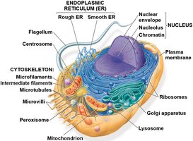
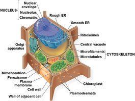
Eukaryotic cells contain various organelles, each with specific functions.
Animal cells: Contain nucleus, ER, Golgi apparatus, mitochondria, lysosomes, cytoskeleton, etc.
Plant cells: Also contain chloroplasts, central vacuole, cell wall, and plasmodesmata.
The Nucleus and Ribosomes
Nucleus: Information Central
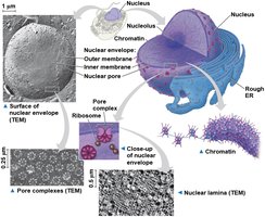
The nucleus houses most of the cell's DNA and is surrounded by a double membrane.
Nuclear envelope: Double membrane with nuclear pores for transport.
Nuclear lamina: Protein network supporting nuclear shape.
Chromatin: DNA-protein complex; condenses into chromosomes during cell division.
Nucleolus: Site of ribosomal RNA synthesis.
Ribosomes: Protein Factories
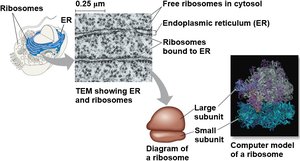
Ribosomes are complexes of RNA and protein, responsible for protein synthesis.
Free ribosomes: In cytosol; synthesize proteins for internal use.
Bound ribosomes: Attached to ER or nuclear envelope; synthesize proteins for secretion or membrane insertion.
The Endomembrane System
Components and Functions
The endomembrane system includes the nuclear envelope, ER, Golgi apparatus, lysosomes, vacuoles, and plasma membrane.
Continuous or connected via vesicles.
Endoplasmic Reticulum (ER)
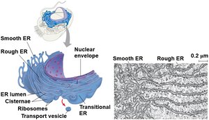
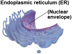
The ER is a network of membranes, continuous with the nuclear envelope.
Smooth ER (SER): Synthesizes lipids, metabolizes carbohydrates, detoxifies, stores calcium.
Rough ER (RER): Studded with ribosomes; synthesizes proteins, distributes vesicles, produces membranes.
Golgi Apparatus
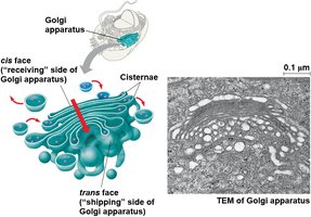
The Golgi apparatus modifies, sorts, and packages proteins and other molecules.
Cisternae: Flattened membranous sacs.
Polarity: Cis (receiving) and trans (shipping) faces.
Lysosomes
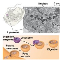
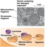
Lysosomes are sacs of hydrolytic enzymes for digestion and recycling.
Phagocytosis: Engulfing and digesting other cells or particles.
Autophagy: Recycling cell's own organelles and macromolecules.
Vacuoles
Vacuoles are large vesicles with diverse functions.
Food vacuoles: Formed by phagocytosis.
Contractile vacuoles: Pump excess water out of cells.
Central vacuole: Stores organic compounds and water in plants.
Mitochondria, Chloroplasts, and Peroxisomes
Energy Conversion Organelles
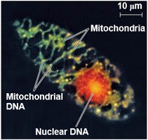
Mitochondria: Sites of cellular respiration; generate ATP.
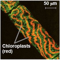
Chloroplasts: Sites of photosynthesis in plants and algae.
Peroxisomes: Oxidative organelles; produce and convert hydrogen peroxide.
Endosymbiont Theory
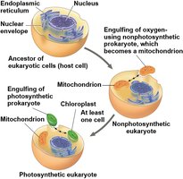
Mitochondria and chloroplasts originated from prokaryotic cells engulfed by ancestral eukaryotes.
Double membrane, free ribosomes, circular DNA.
Grow and reproduce independently within cells.
Mitochondria Structure
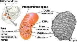
Mitochondria have an outer and inner membrane, with the inner membrane folded into cristae.
Matrix: Contains enzymes for cellular respiration.
Cristae: Increase surface area for ATP synthesis.
Chloroplast Structure
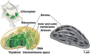
Chloroplasts contain thylakoids, stacked into grana, and stroma.
Thylakoids: Membranous sacs for light reactions.
Stroma: Internal fluid for Calvin cycle.
The Cytoskeleton
Structure and Function
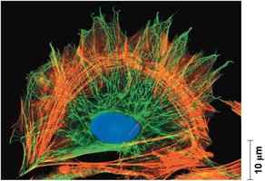
The cytoskeleton is a network of fibers that organizes cell structure and activities.
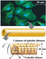
Microtubules: Thickest, made of tubulin; shape, organelle movement, chromosome separation.
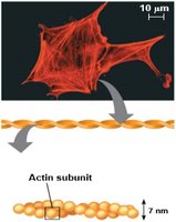
Microfilaments (Actin): Thinnest; support cell shape, motility, muscle contraction.
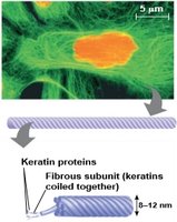
Intermediate Filaments: Middle diameter; maintain cell shape, anchor organelles.
Motor Proteins and Cell Motility
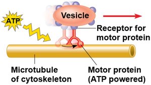
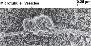
Motor proteins interact with cytoskeletal fibers to move vesicles and organelles.
ATP-powered movement: Motor proteins "walk" along microtubules.
Microtubules, Microfilaments, and Intermediate Filaments
Each cytoskeletal component has unique structure and function.
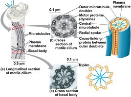
Microtubules: Hollow rods, 25 nm diameter; centrosomes, centrioles, cilia, flagella.
Microfilaments: Twisted double chain of actin; cortex, microvilli, muscle contraction, cell motility.
Intermediate Filaments: 8–12 nm diameter; keratin, structural support.
Centrosomes and Centrioles
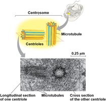
Microtubules grow from centrosomes, which contain centrioles.
Centrioles: Nine triplets of microtubules arranged in a ring.
Cilia and Flagella
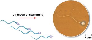
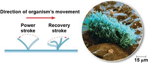
Cilia and flagella are microtubule-based structures for cell movement.
Cilia: Short, numerous, move fluid over cell surface.
Flagella: Long, few, propel cells.
Microfilament Functions
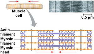
Microfilaments support cell shape and enable motility.
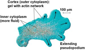
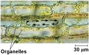
Actin-myosin interactions: Muscle contraction, cytoplasmic streaming, amoeboid movement.
Extracellular Components and Cell Junctions
Cell Walls of Plants
Plant cells have cell walls made of cellulose, providing protection and structural support.
Primary cell wall: Thin and flexible.
Middle lamella: Layer between adjacent cells.
Secondary cell wall: Added for extra strength.
Extracellular Matrix (ECM) of Animal Cells
Animal cells are surrounded by ECM, composed of glycoproteins like collagen and proteoglycans.
Integrins: ECM proteins bind to cell surface receptors, influencing cell behavior and gene expression.
Cell Junctions
Cells communicate and adhere through specialized junctions.
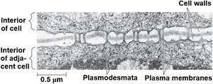
Plasmodesmata: Channels in plant cell walls for transport of water and solutes.
Tight junctions: Prevent leakage between animal cells.
Desmosomes: Anchor cells together.
Gap junctions: Allow communication between animal cells.
Integration of Cell Structure and Function
Coordination of Cellular Activities
Cells rely on the integration of organelles and structures to perform complex functions, such as immune responses and metabolism.
Example: Macrophages use cytoskeleton, lysosomes, and plasma membrane to destroy bacteria.
Summary Tables
Nucleus and Ribosomes
Cell Component | Structure | Function |
|---|---|---|
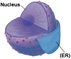
Nucleus | Surrounded by nuclear envelope (double membrane) with pores; continuous with ER | Houses chromosomes (chromatin); contains nucleoli; regulates entry/exit of materials |
Ribosomes | Two subunits made of rRNA and proteins; free in cytosol or bound to ER | Protein synthesis |
Endomembrane System
Cell Component | Structure | Function |
|---|---|---|
ER | Network of membrane-bounded tubules and sacs; continuous with nuclear envelope | Smooth ER: lipid synthesis, carbohydrate metabolism, storage, detoxification Rough ER: protein synthesis, glycoprotein formation, membrane production |
Golgi Apparatus | Stacks of flattened sacs; cis and trans faces | Modification, sorting, packaging of proteins and lipids |
Lysosome | Membranous sac of hydrolytic enzymes | Breakdown of substances, recycling |
Vacuole | Large membrane-bounded vesicle | Digestion, storage, waste disposal, water balance, cell growth, protection |
Mitochondria, Chloroplasts, and Peroxisomes
Cell Component | Structure | Function |
|---|---|---|
Mitochondria | Double membrane; inner membrane with infoldings | Cellular respiration |
Chloroplast | Two membranes around stroma; thylakoids stacked into grana | Photosynthesis |
Peroxisome | Single membrane | Oxidation reactions; hydrogen peroxide production and conversion |
