 Back
A Tour of the Cell: Structure, Function, and Organization
Study Guide - Smart Notes
 Tailored notes based on your materials, expanded with key definitions, examples, and context.
Chapter 4: A Tour of the Cell
Introduction to Cell Structure and Function
Cells are the fundamental units of life, and their internal organization allows them to perform the essential functions required for life. This chapter explores the diversity of cell types, the tools used to study them, and the structural features that distinguish prokaryotic and eukaryotic cells.
Microscopy and Cell Study
Microscopy: Tools for Cell Biology
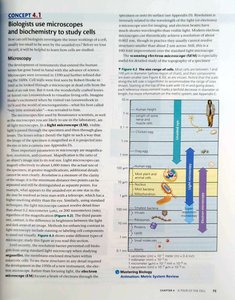
Microscopes are essential for studying cells, which are typically too small to be seen with the naked eye. There are several types of microscopes, each with unique capabilities:
Light Microscopes (LM): Use visible light to illuminate specimens. Useful for viewing living cells and tissues up to about 1000x magnification.
Electron Microscopes (EM): Use beams of electrons for much higher resolution. Includes Transmission Electron Microscopes (TEM) for internal structures and Scanning Electron Microscopes (SEM) for surface details.
Cell Fractionation: A technique to separate cellular components for biochemical analysis.
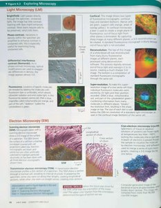
Types of Microscopy
Brightfield (unstained and stained): Standard light microscopy, sometimes with dyes to enhance contrast.
Phase-contrast and Differential Interference Contrast (DIC): Enhance contrast in unstained cells.
Fluorescence Microscopy: Uses fluorescent dyes or proteins to label specific cell components.
Confocal Microscopy: Provides optical sectioning for clearer images of thick specimens.
Electron Microscopy: Reveals ultrastructure, such as organelles and macromolecular complexes.
Cell Types and Internal Organization
Prokaryotic vs. Eukaryotic Cells
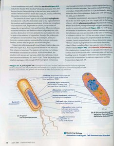
Cells are classified as prokaryotic or eukaryotic based on their internal structure:
Prokaryotic Cells: Lack a nucleus and membrane-bound organelles. DNA is located in the nucleoid region. Examples: Bacteria and Archaea.
Eukaryotic Cells: Have a true nucleus enclosed by a nuclear envelope and numerous membrane-bound organelles. Examples: Plants, animals, fungi, and protists.
Cell Membranes and Compartmentalization
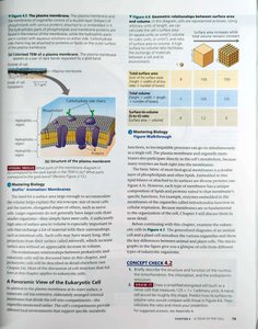
The plasma membrane defines the boundary of the cell and regulates the movement of substances in and out. Eukaryotic cells have internal membranes that compartmentalize functions, allowing for specialization and efficiency.
Surface Area to Volume Ratio: Limits cell size; as a cell grows, its volume increases faster than its surface area, affecting material exchange.
Overview of Eukaryotic Cell Structure
Animal and Plant Cell Structures
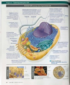
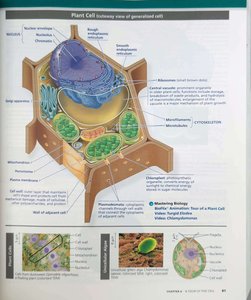
Eukaryotic cells contain a variety of organelles, each with specialized functions:
Nucleus: Contains genetic material (DNA) and controls cellular activities.
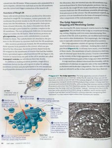
Endoplasmic Reticulum (ER): Rough ER synthesizes proteins; Smooth ER synthesizes lipids and detoxifies chemicals.
Golgi Apparatus: Modifies, sorts, and packages proteins and lipids for transport.
Mitochondria: Sites of cellular respiration and energy (ATP) production.
Chloroplasts (plants only): Sites of photosynthesis.
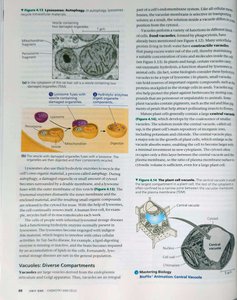
Lysosomes: Contain digestive enzymes for breaking down macromolecules.
Vacuoles: Storage and structural support (large central vacuole in plants).
Cytoskeleton: Network of protein fibers for support, movement, and transport.
The Nucleus and Ribosomes
Nucleus: Information Central
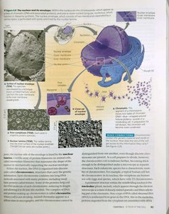
The nucleus is surrounded by a double membrane (nuclear envelope) and contains most of the cell's DNA. The nucleolus within the nucleus is the site of ribosomal RNA (rRNA) synthesis and ribosome assembly.
Nuclear Pores: Regulate entry and exit of molecules.
Chromatin: DNA and proteins that condense to form chromosomes during cell division.
Ribosomes: Protein Factories
Ribosomes are complexes of rRNA and protein that carry out protein synthesis. They can be free in the cytosol or bound to the rough ER.
The Endomembrane System
Components and Functions
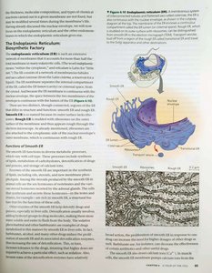
The endomembrane system includes the nuclear envelope, ER, Golgi apparatus, lysosomes, vacuoles, and plasma membrane. It regulates protein traffic and performs metabolic functions.
Rough ER: Studded with ribosomes; synthesizes proteins for secretion or membrane insertion.
Smooth ER: Lacks ribosomes; synthesizes lipids, metabolizes carbohydrates, detoxifies drugs and poisons, stores calcium ions.
Golgi Apparatus: Receives, modifies, and ships products from the ER.
Lysosomes: Digestive organelles where macromolecules are hydrolyzed.
Vacuoles: Diverse functions including storage, waste disposal, and maintaining turgor pressure in plants.
Mitochondria and Chloroplasts: Energy Conversion
Mitochondria
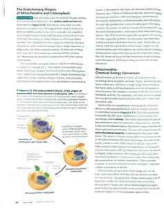
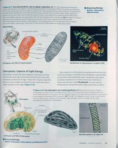
Mitochondria are the sites of cellular respiration, converting glucose and oxygen into ATP, the cell's energy currency. They have a double membrane and their own DNA, supporting the endosymbiont theory of origin.
Chloroplasts
Chloroplasts are found in plants and algae and are the sites of photosynthesis, converting light energy into chemical energy stored in glucose. They also have a double membrane and their own DNA.
Other Organelles and Structures
Peroxisomes
Peroxisomes are small organelles that contain enzymes for oxidation reactions, breaking down fatty acids and detoxifying harmful substances.
The Cytoskeleton
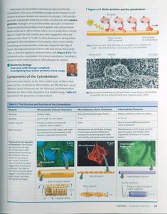
The cytoskeleton is a network of protein fibers that provides structural support, facilitates cell movement, and organizes cellular components. It includes:
Microtubules: Hollow rods that shape the cell, guide organelle movement, and separate chromosomes during cell division.
Microfilaments (Actin Filaments): Thin rods that support the cell's shape and are involved in movement.
Intermediate Filaments: Provide mechanical support for the cell.
Cilia and Flagella
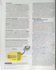
Cilia and flagella are motile structures composed of microtubules, enabling movement of cells or movement of substances along cell surfaces.
Extracellular Components and Cell Junctions
Plant Cell Walls
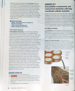
Plant cells are surrounded by a rigid cell wall made of cellulose, providing structural support and protection.
Extracellular Matrix (ECM) of Animal Cells
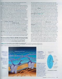
The ECM is a network of glycoproteins and other molecules outside animal cells that provides structural support, segregates tissues, and regulates intercellular communication.
Cell Junctions
Cells communicate and adhere to each other through specialized junctions:
Tight Junctions: Prevent leakage of extracellular fluid.
Desmosomes: Anchor cells together.
Gap Junctions: Allow passage of ions and small molecules between cells.
Plasmodesmata (plants): Channels that connect plant cells for transport and communication.
Summary Table: Major Differences Between Prokaryotic and Eukaryotic Cells
Feature | Prokaryotic Cells | Eukaryotic Cells |
|---|---|---|
Nucleus | Absent | Present |
Membrane-bound Organelles | Absent | Present |
Size | 1–10 μm | 10–100 μm |
Examples | Bacteria, Archaea | Plants, Animals, Fungi, Protists |
Key Equations
Surface Area of a Sphere:
Volume of a Sphere:
Surface Area to Volume Ratio:
Additional info: This summary integrates and expands upon the provided textbook pages, ensuring a comprehensive overview of cell structure and function for introductory biology students.
