 Back
A Tour of the Cell: Structure, Function, and Tools of Cytology
Study Guide - Smart Notes
 Tailored notes based on your materials, expanded with key definitions, examples, and context.
Tools of Cytology
Microscopy: Revealing the Invisible World of Cells
Cells are the fundamental units of life, but their internal structures are too small to be seen by the unaided eye. Biologists use various types of microscopes to visualize and study cells and their components.
Magnification: The ratio of an object's image size to its real size.
Resolution: The clarity of the image, or the minimum distance between two distinguishable points.
Contrast: The difference in brightness between the light and dark areas of an image.
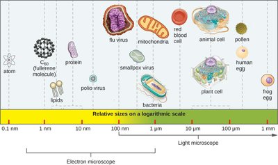
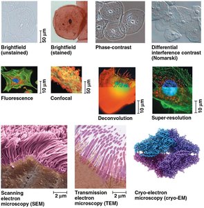
Light microscopes (LM) use visible light and are limited by its wavelength, while electron microscopes (EM) use electron beams with much shorter wavelengths, allowing for higher resolution and visualization of organelle ultrastructure.
Cell Fractionation: Isolating Cellular Components
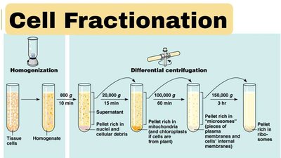
Cell fractionation is a technique that separates cellular components based on size and density using differential centrifugation. This allows researchers to study the function of individual organelles in isolation.
Process: Cells are homogenized and spun at increasing speeds. Larger components (nuclei) settle first, followed by mitochondria, then ribosomes.
Application: Enables the study of biochemical activities of specific organelles.
Cellular Organization and Compartmentalization
Prokaryotic vs. Eukaryotic Cells
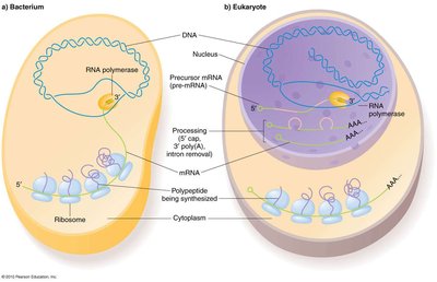
All cells share a plasma membrane, cytosol, and ribosomes. However, eukaryotic cells possess a nucleus and extensive internal membranes, while prokaryotes lack these features.
Prokaryotes: Simpler structure, DNA in a nucleoid region, no membrane-bound organelles.
Eukaryotes: DNA enclosed in a nucleus, compartmentalization via organelles, greater complexity.
Surface Area-to-Volume Ratio
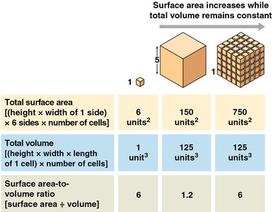
The surface area-to-volume ratio limits cell size. As a cell grows, its volume increases faster than its surface area, restricting efficient exchange of materials with the environment.
Implication: Cells remain small or adopt shapes that maximize surface area for metabolic efficiency.
Cytosol vs. Organelles
Cytosol is the liquid matrix of the cytoplasm, supporting biochemical reactions and organelles. Organelles are specialized structures performing distinct cellular functions (e.g., mitochondria, ER, Golgi apparatus).
Information Storage and Flow
The Nucleus: Genetic Archive
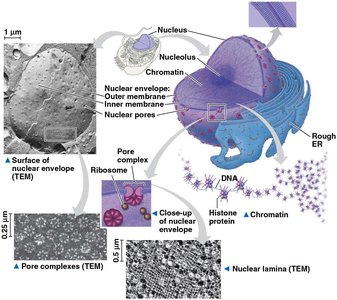
The nucleus houses the cell's genetic material within a double-membrane envelope. Nuclear pores regulate the exchange of materials, and the nucleolus synthesizes ribosomal RNA (rRNA).
Chromatin: DNA-protein complex that condenses into chromosomes during cell division.
Nucleolus: Site of rRNA synthesis and ribosome assembly.
Endoplasmic Reticulum (ER): Biosynthetic Factory
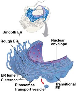
The ER is a network of membranes continuous with the nuclear envelope. It has two regions:
Smooth ER: Lacks ribosomes; involved in lipid synthesis, carbohydrate metabolism, and detoxification.
Rough ER: Studded with ribosomes; synthesizes proteins for membranes, organelles, or secretion.
Ribosomes: Protein Synthesis Machinery
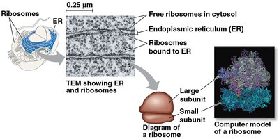
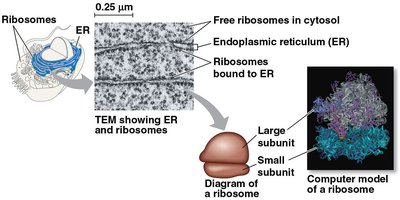
Ribosomes are complexes of rRNA and protein that translate mRNA into polypeptides. They exist as free ribosomes (cytosol) or bound ribosomes (ER).
Free ribosomes: Synthesize proteins for use within the cytosol.
Bound ribosomes: Synthesize proteins for membranes, organelles, or secretion.
Golgi Apparatus: Shipping and Receiving Center
The Golgi apparatus consists of flattened sacs (cisternae) that modify, sort, and package proteins and lipids from the ER. It adds molecular tags to direct products to their destinations.
Cis face: Receiving side.
Trans face: Shipping side.
Energy Converters
Mitochondria and Chloroplasts: Endosymbiont Theory
Mitochondria and chloroplasts are energy-converting organelles believed to have originated from prokaryotic ancestors via endosymbiosis. Both have double membranes, their own DNA, and replicate independently.
Mitochondria: Site of cellular respiration, converting chemical energy into ATP.
Chloroplasts: Site of photosynthesis, converting light energy into chemical energy (sugars).
Peroxisomes: Detoxification Compartments
Peroxisomes contain enzymes that transfer hydrogen from substrates to oxygen, producing hydrogen peroxide (H2O2), which is then converted to water. They detoxify harmful substances and break down fatty acids.
Lysosomes: Waste Management
Lysosomes are membrane-bound organelles containing hydrolytic enzymes for digesting macromolecules, recycling cellular components (autophagy), and destroying pathogens (phagocytosis). Lysosomal failure can lead to diseases such as Tay-Sachs.
The Cytoskeleton: Cellular Framework
Types of Cytoskeletal Fibers
Microtubules: Hollow rods of tubulin; provide tracks for organelle movement and chromosome separation during cell division.
Microfilaments (Actin Filaments): Twisted chains of actin; support cell shape, enable muscle contraction, and drive cell motility.
Intermediate Filaments: Cable-like proteins; provide permanent structural support and anchor organelles.
Specialized Structures: Cilia, Flagella, and Centrosomes
Cilia and Flagella: Microtubule-based extensions for cell movement; share a '9+2' arrangement of microtubules.
Centrosomes and Centrioles: Organize microtubule growth in animal cells.
Motor Proteins
Motor proteins use ATP to "walk" vesicles and organelles along cytoskeletal tracks, enabling directed transport within the cell.
Extracellular Coordination and Cell Communication
Extracellular Matrix (ECM) in Animal Cells
The ECM is a network of glycoproteins (e.g., collagen, fibronectin) that provides structural support and mediates communication between cells via integrin receptors.
Cell Junctions
Tight Junctions: Seal cells together to prevent leakage.
Desmosomes: Fasten cells into strong sheets.
Gap Junctions: Provide cytoplasmic channels for communication.
Plasmodesmata (plants): Channels that connect plant cells for solute exchange.
Plant Cell Walls
Plant cell walls are composed of cellulose fibers, providing protection, shape, and preventing excessive water uptake. The middle lamella glues adjacent cells together.
Prokaryotic Cell Structure
Prokaryotic cells lack compartmentalization but efficiently carry out all necessary life processes. Their DNA is located in a nucleoid region, and they possess structures such as fimbriae, flagella, and a glycocalyx for attachment and protection.
Vacuoles and Plastids
Vacuoles
Vacuoles are membrane-bound compartments with diverse functions:
Plant cells: Large central vacuole for storage, turgor pressure, and structural support.
Animal cells: Small, numerous vacuoles for waste sequestration and endocytosis.
Fungal cells: Acidic storage compartments for ion/nutrient storage and degradation.
Plastids
Plastids are specialized organelles in plant cells, including:
Amyloplasts: Store starch.
Chromoplasts: Synthesize and store pigments.
Leucoplasts: Involved in storage and biosynthesis of compounds.
Summary Tables
Microscopy and Cell Fractionation
Process | Component | Function |
|---|---|---|
Microscopy (LM vs. EM) | Visualization | Resolving subcellular structures |
Differential Centrifugation | Physical Separation | Isolating organelles for functional study |
Cellular Organization
Process | Component | Function |
|---|---|---|
Compartmentalization | Internal Membranes | Separating incompatible metabolic reactions |
Metabolic Exchange | Plasma Membrane | Regulating passage of matter relative to volume |
Information Flow
Process | Component | Function |
|---|---|---|
Genetic Storage | Chromatin / Nucleus | Protecting the hereditary archive |
Structural Synthesis | Nucleolus | Building ribosomal subunits |
Translation | Ribosomes | Converting mRNA into polypeptides |
Energy Converters
Process | Component | Function |
|---|---|---|
Cellular Respiration | Mitochondria | Transforming chemical energy into ATP |
Photosynthesis | Chloroplasts | Transforming light energy into chemical energy |
Detoxification | Peroxisomes | Breaking down fatty acids and toxins |
The Cell's Skeleton
Process | Component | Function |
|---|---|---|
Track Guidance | Microtubules | Moving organelles and chromosomes |
Contraction / Support | Microfilaments (Actin) | Muscle movement and cell shape (cortex) |
Permanence | Intermediate Filaments | Fixing the nucleus in place |
Extracellular Matrix and Cellular Integration
Process | Component | Function |
|---|---|---|
Architectural Support | Glycoproteins (Collagen) | Providing a structural framework for animal tissues |
Molecular Anchorage | Fibronectin and Integrins | Linking the extracellular environment to the internal cytoskeleton |
Functional Regulation | Integrin Signaling | Influencing nuclear gene expression based on environmental cues |
Process | Component | Function |
|---|---|---|
Structural Barrier | Cell Walls / Tight Junctions | Protecting and sealing tissues |
External Signaling | Extracellular Matrix (ECM) | Communicating with the nucleus (integrins) |
Direct Communication | Gap Junctions / Plasmodesmata | Exchanging solutes between neighbors |
