 Back
A Tour of the Cell: Structure and Function in Eukaryotic and Prokaryotic Cells
Study Guide - Smart Notes
 Tailored notes based on your materials, expanded with key definitions, examples, and context.
The Cell: Basic Unit of Structure and Function
Overview of Cellular Organization
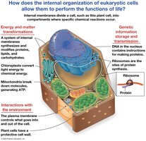
The cell is the fundamental unit of life, providing structure and function for all living organisms. Eukaryotic cells possess internal membranes that compartmentalize their functions, allowing specialized chemical reactions to occur efficiently.
Energy and Material Transformation: Organelles such as mitochondria and chloroplasts convert energy and synthesize essential molecules.
Genetic Information Storage and Transmission: The nucleus stores DNA, which is transcribed and translated by ribosomes to produce proteins.
Interactions with the Environment: The plasma membrane regulates the exchange of substances and signals with the environment.
Microscopy: Observing Cells
Principles and Types of Microscopy
Microscopy enables the observation of cells and their structures, which are too small to be seen with the naked eye.
Magnification: The ratio of an object's image size to its real size. Formula:
Resolution: The clarity of the image, or the minimum distance between two distinguishable points.
Contrast: The difference in brightness between light and dark areas of the image.
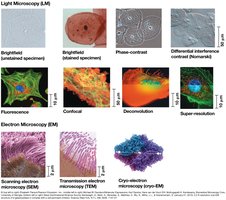
Light microscopes (LM) can magnify up to 1,000 times, but most organelles are too small to be resolved. Electron microscopes (EM) provide higher resolution, allowing visualization of subcellular structures. 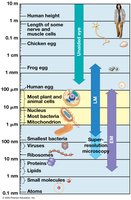
Types of Cells: Prokaryotic vs. Eukaryotic
Cell Classification and Domains of Life
All organisms are classified into three domains: Bacteria, Archaea, and Eukaryotes.
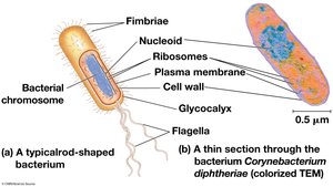
Prokaryotic Cells: Found in Bacteria and Archaea. Lack a nucleus and membrane-bound organelles. DNA is located in the nucleoid region. Size: 1-5 µm.
Eukaryotic Cells: Found in protists, fungi, animals, and plants. Have a nucleus and membrane-bound organelles. Size: 10-100 µm.
Basic Features of All Cells
Plasma membrane: Selective barrier for transport.
Cytosol: Semifluid substance inside the cell.
Chromosomes: Carry genetic information.
Ribosomes: Sites of protein synthesis.
Eukaryotic Cell Structure
Animal and Plant Cell Organization
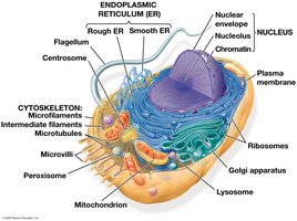
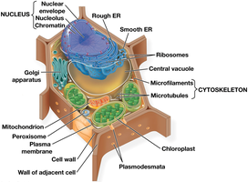
Eukaryotic cells contain extensive internal membranes and organelles, each with specialized functions.
Animal Cells: Contain nucleus, endoplasmic reticulum, Golgi apparatus, mitochondria, lysosomes, peroxisomes, cytoskeleton, and plasma membrane.
Plant Cells: Contain all animal cell organelles plus chloroplasts, central vacuole, cell wall, and plasmodesmata.
The Nucleus: Information Central
Structure and Function of the Nucleus
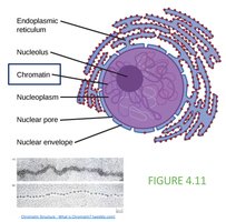
The nucleus is the most prominent organelle, containing most of the cell's genetic material.
Nuclear Envelope: Double membrane with nuclear pores for molecular transport.
Chromatin: DNA and associated proteins, organized into chromosomes.
Nucleolus: Site of ribosomal RNA (rRNA) synthesis.
Ribosomes: Protein Factories
Structure and Function of Ribosomes
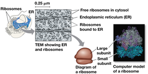
Ribosomes are complexes of rRNA and protein, responsible for protein synthesis.
Free Ribosomes: Located in the cytosol; synthesize proteins for use within the cell.
Bound Ribosomes: Attached to the endoplasmic reticulum or nuclear envelope; synthesize proteins for secretion or membrane insertion.
The Plasma Membrane
Structure and Function
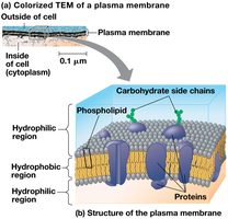
The plasma membrane is a selective barrier composed of a phospholipid bilayer with embedded proteins. It regulates the passage of oxygen, nutrients, and waste.
Phospholipid Bilayer: Hydrophilic heads face outward, hydrophobic tails face inward.
Proteins: Serve as channels, receptors, and enzymes.
The Endomembrane System
Components and Functions
The endomembrane system includes the nuclear envelope, endoplasmic reticulum, Golgi apparatus, lysosomes, vesicles, vacuoles, and plasma membrane. These components are interconnected and regulate protein traffic and metabolic functions.
The Endoplasmic Reticulum (ER)
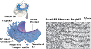
Smooth ER: Synthesizes lipids, metabolizes carbohydrates, detoxifies drugs.
Rough ER: Studded with ribosomes; synthesizes glycoproteins, distributes transport vesicles, and produces membrane.
The Golgi Apparatus
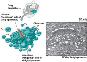
Structure: Flattened membranous sacs called cisternae.
Function: Modifies, sorts, and packages products from the ER into transport vesicles.
Lysosomes
Structure: Membranous sac of hydrolytic enzymes.
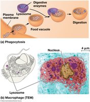
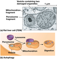
Function: Digests macromolecules, recycles organelles (autophagy), and participates in phagocytosis.
Vacuoles
Food Vacuoles: Formed by phagocytosis.
Contractile Vacuoles: Pump excess water out of cells (in protists).
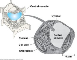
Central Vacuole: Found in plant cells; stores water, nutrients, and maintains turgor pressure.
Mitochondria and Chloroplasts: Energy Conversion
Mitochondria
Mitochondria are the sites of cellular respiration, generating ATP from the breakdown of sugars and fats.
Structure: Double membrane, inner membrane folded into cristae, matrix, and intermembrane space.
Function: Catalyzes metabolic steps of cellular respiration.
Chloroplasts
Chloroplasts are the sites of photosynthesis in plants and algae.
Structure: Contains thylakoids (stacked as granum), stroma, and chlorophyll.
Function: Converts solar energy to chemical energy (sugars).
Endosymbiont Theory
Mitochondria and chloroplasts are believed to have originated from prokaryotic cells engulfed by ancestral eukaryotes.
Evidence: Double membranes, own ribosomes and circular DNA, independent replication.
The Cytoskeleton: Support and Motility
Structure and Function
The cytoskeleton is a network of fibers that organizes cell structures and activities.
Microtubules: Thickest fibers; shape, support, guide organelle movement, and separate chromosomes.
Microfilaments (Actin Filaments): Thinnest fibers; bear tension and form microvilli.
Intermediate Filaments: Middle diameter; provide structural support.
Specialized Structures
Centrosomes and Centrioles: Microtubule-organizing centers in animal cells.
Cilia and Flagella: Motile structures controlled by microtubules and motor proteins.
Extracellular Components and Cell Junctions
Cell Walls in Plants
The cell wall provides protection, shape, and prevents excessive water uptake.
Primary Cell Wall: Thin and flexible.
Middle Lamella: Layer between adjacent cells.
Secondary Cell Wall: Added in some cells for extra strength.
Extracellular Matrix (ECM) in Animals
Animal cells are surrounded by ECM, composed of glycoproteins like collagen, proteoglycans, and fibronectin.
Cell Junctions
Plasmodesmata: Channels in plant cell walls for transport and communication.
Tight Junctions, Desmosomes, Gap Junctions: Specialized junctions in animal cells for adhesion and communication.
Integration of Cellular Components
Coordination of Cell Functions
Cellular components work together to perform complex functions, such as immune responses, metabolism, and communication.
Example: Macrophages coordinate cytoskeleton, lysosomes, and plasma membrane to destroy bacteria.
Additional info: This study guide covers the main concepts from Chapter 4: A Tour of the Cell, including cell structure, microscopy, organelles, and cellular functions, as outlined in a General Biology college course.
